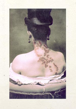

Endurance & Suffering has won the Deutscher Fotobuchpreis for 2009, taking the gold medal for Historical—Theoretical Photography! Heartfelt congratulations to the poet John Wood and his publisher, Alexander Scholz! !!!
Albert Buck's paper on a pathological specimen from a child's inner ear, prepared by the great Julius Arnold of Heidelberg in 1869. He was the son of Dr. Gurdon Buck.
James Anderson, M. D., Chairman, &c.—
Dear Sir: As Chairman of the Committee of the American Medical Association having charge of such matters, I beg
leave to request through you the privilege of presenting to that body, at such time as they may see fit to appoint,
a case in which an extensive destruction of the face by cancrum oris has been successfully repaired by a series of
plastic operations. The patient himself will be exhibited, as well as various plastic masks and photographic drawings
illustrating the several operations. The extraordinary interest possessed by this unique case, it is believed, will
warrant the undersigned in respectfully soliciting the indulgence of the Association for one hour for the purpose of
exhibiting it to their notice.
Awaiting a reply at your earliest convenience, I remain,
Very respectfully yours, &c., GURDON BUCK
On motion, Dr. Gurdon Buck, of New York, was requested to introduce a patient who had been operated on for destruction of the right side of his face by a number of successive plastic operations. Dr. Buck referred the members to the minute description of the case, which will be published in the Transactions of the New York State Medical Society for 1864, and explained the different steps of the operation briefly, illustrating his remarks with photographs and casts taken at different periods.
Cataloging resumes with this paper on the morphology of the inner ear of an elephant.
Update begun on the Randall & Morse photographic atlas of the ear.
In pursuance of a subject which Dr. Morse and I brought before this Society two years ago, I beg leave to show some more photographic illustrations ; but to present them this time in the form of lantern slides to indicate how excellently photography may be made to serve us by the use of projections upon the screen in teaching our specialty. I have not limited the series now to the anatomv of the human ear and the pathology of the drum-membrane ; but include clinical pictures and selections from preparations in comparative anatomy. Among the latter I may call attention to the preparations made by Hyrtl of the tympanic membrane, ossicles, and labyrinth of the elephant as of interest in connection with the demonstrations made here last year by Dr. Buck. As has been before stated, these plates are entirely free from retouching ; so while their beauty may be less than skillful retouching would secure, they are unimpeachable in their accuracv. They have already served me well with my classes at the Philadelphia Polyclinic, and I propose to still further extend the series of which these form a part. — Page 484, vol. iv, Transactions of the American Otological Society (1887).
A photo of a specimen of a cyclops illustrates this paper. It is strange how a piece of a body, especially an eye, can seem to be infused with the presence of an intelligence. Even the dumb eye of a dead cyclops evokes the single polarity of conciousness. Gather together enough of these sticks and you have an organism reading the morning newspaper and ordering cappucino at a diner.
A paper on examination of the eye.
Paper on a thoracopagus monster.
Interesting neurological experiment for which the phrenic nerve was cut in rabbits.
Updated Dean.
Added a paper by Thomas R. French on photographing the larynx.
Added a paper by Ephraim Cutter on photographing the larynx.
Began cataloging a paper by a once beloved Brooklyn surgeon for the placement of the first prosthetic larynx.
Since the report which he had made at the last meeting a large number of experiments had been tried, and various improvements been made in taking photographs of the larynx, in all of which he had been enthusiastically seconded by his friend, Mr. Brainard, of Brooklyn, a civil engineer by profession, and an accomplished amateur photographer. Their aim had been, first, to simplify the procedure and render it of greater practical utility, and, secondly, to make better photographs. Of late a hand camera, instead of the stationary instrument formerly employed, had been tried, and although the pictures taken by it were considerably smaller in size it rendered it possible to secure photographs of many patients, in whom the larynx permitted only a moderate degree of tolerance, which could not be taken with the stationary camera. The light employed had been of five kinds, namely, unaided sunlight, condensed sunlight, and the oxy-hydrogen, magnesium, and electric lights. The electric light, he said, had been found much less efficient than condensed sunlight, which had proved to be the most satisfactory kind of illumination. While he had most commonly employed the hand camera of late he had not abandoned the use of the stationary instrument in suitable cases, and the photographs taken with it during the last few months were much better than those of last year. Still they were not of much practical value.
Dr. French then described the hand camera and its appendages, including a condenser of sunlight, devised by Mr. Brainard. Some of the photographs (a number of which were exhibited), he said, were so small that they could not be seen well without a magnifying glass, and in order to make them fully appreciable by the unaided eye it was necessary to enlarge them. By the use of more perfect lenses and the adoption of other improvements he hoped to accomplish better results in the future than had yet been attained. The results achieved since last year were summed up as follows: —
(1.) Better photographs taken with the stationary camera. (2.) The camera so modified as to be held in the hand. (3.) Photographs taken instantaneously by means of the drop-shutter. (4.) The parts exposed in the mirror alone photographed. (5.) The securing of photographs without the knowledge of the patient when desirable. (6.) Several diseased conditions of the larynx photographed for the first time. (7.) Portions of the rhinoscopic image taken for the first time. — BMSJ May 31, 1883 (p. 518).
A neurological riddle from 1889.
William Noyes claimed that he made first composite photographs of the insane.
Textbook of Ranney who built a thriving practice treating New York neurotics with electric shock and eye surgery.
December 7.
WONDERS OF THE CAMERA. — The peculiar rhythmical effects which accompany discharges of powder and of nitroglycerine compounds have been elaborately investigated by the aid of photography. It has also been suggested that careful photographs, taken of steel and timber just at the point of rupture under a breaking load, would conduce to our knowledge of the complicated subject of elasticity. The lightning flash can be investigated. Dr. Koenig, in a recent communication to the Physical Society of Berlin, states that he has photographed a cannon-ball which was moving at a rate of 1200 feet per second. The ball was projected in front of a white screen and occupied one fortieth of a second in its passage. Marey has photographed the motions of limping people, and has thus given surgeons the materials for a study of lameness. It is said, moreover, that photography often reveals incipient eruptive diseases which are not visible to the eye. Photographs taken by flash-powders of the human eye, showing it dilated in the dark, give the oculist a new method of studying the enlarged pupil. — Professor John Trowbridge, in the May Scribner's (extract from The Sanitarian, vol. xxii, 1889).
Pregnancy in a woman with a deformed pelvis.
Finished cataloging Smith's paper on Friedreich's ataxia. The latest sumptuous Alain Brieux catalog is out with first edition offerings of the Hardy & Montmeja atlas and Duchenne's 1862 masterpiece on physiognomy.
Cataloging begun for a paper on Friedreich's ataxia. Illustrated by 2 crappy half-tones.
Paper on the spontaneous breakup of urinary stones. Illustrated by a crappy half-tone.
Paper on the first gastrotomy for removal of a foreign object lodged in the esophagus. Illustrated by a crappy half-tone.
Began cataloging a case report by Hack Tuke.
Note to self:
The observation of the insane is a
fruitful source of knowledge, and I have during the last
ten years obtained a large number of photographs from
various institutions, more especially the Cambridge Asylum,
Earlswood, Essex Hall, and Bethlem Hospital. I am
particularly indebted to Dr Savage for a valuable series of
photographs of patients, taken at the last mentioned
asylum. I have, of course, availed myself of the opportunity
of observing the expressions of patients themselves
in a variety of mental states, and this in many instances is
more trustworthy than photographs, because the attention
of the patient is in the latter often arrested and diverted
into a fresh channel of thought. — Illustrations of the Influence of
the Mind Upon the Body in Health and Disease, By Daniel Hack Tuke (page 229).
Paper on the criminally insane by the deputy superintendent of Broadmoor asylum. By 1895 there were 650 men locked up in the institution.
Paper on a xiphopagus monster.
Revilliod's paper on intubation of the liver.
Reverdin's paper on amputation at the hip-joint.
Griesinger's contribution to the literature on myopathy.
First reports from the Hospital de Clinicas in Buenos Aires.
This offprint by an Italian inventor is the first published photograph of a vaporizor.
A study on sexual perversion, extraordinary for its photographic illustration.
One more doctoral thesis from Buenos Aires.
A treatise on knee surgery by a Genova surgeon.
Another doctoral thesis from Buenos Aires.
A revised anatomical study by Jules Bernard Luys on the subthalamic nucleus which bears his name.
Blanche Dumas reappears in medical literature with this paper by Dr. Bechtinger.
Added a narrative on the photographing in a leper colony.
A second paper by the surgeon Edward H. Bennett — on plastic restoration of the nose.
A paper on hip-joint disease by a master orthopedic surgeon.
Case of lymphadenoma in the neck. Pirovano's delicate operation using a surgical spatula to suppress hemorrhage.
A paper on ichthyosis.
Possibly the first photograph published in Argentina.
A third albumen photograph from volume xiv of the Revista médico-quirúrgica, this time showing a massive lipoma.
Another albumen photograph from volume xiv of the Revista médico-quirúrgica.
Completed cataloging Coni.
Completed cataloging the first of two Emilio Coni's works.
Began cataloging the great Emilio Coni.
Here is the doctoral thesis of one of Argentina's most accomplished physician/scholars.
Surgical and medical case reports for the year 1878 at the Hospital de Niños in Buenos Aires.
This doctoral thesis is illustrated with four albumens representing a case of mandibular osteosarcoma in a 7 year-old child.
Not in the Margolis and Moss catalog, but here is another doctoral thesis, this one on tapeworm.
Began cataloging a doctoral dissertation on one of the first hyperbaric chambers.
This monograph on skull shaping probably belongs in a bibliography on ethnography, but it is included in the Index Medicus so it makes the cut for our bibliography. Also, Lenhossék included a report and illustration of a living child with an unusual saddle-shaped skull. The next several days will be devoted to researching and cataloging the splendid medical offerings in the latest Margolis and Moss catalog.
A tract on the curative powers of the mineral water at Saxon-les-Bains.
Another doctoral dissertation — on a condition with an unknown pathogenesis.
Here is an early atlas of histology with beautiful plates created by Josef Albert.
Worldcat retrieves only one copy of this dissertation on purulent fistules.
Pierre Marie's brilliant work in describing the pituitary disorder of acromegaly. One of the most stunning photographic portraits of the nineteenth century is plate 49, representing the widow Beaufils who suffered from this condition.
A case of tabetic arthropathy in the foot.
A case of unilateral lentigo in an epileptic.
Collotypes illustrating a case of sporadic cretinism.
Catalogued this excerpt from a discourse among French alienists trying to sort out the terminology of cognitive disorders.
Brain specimen showing ventriculomegaly and vestigial corpus callosum in a case of idiocy.
Recommencing the bibliographical work with this paper by the Paris surgeon, Octave Terrillon.
Work on the Cabinet bibliography is postponed until a paper on the Bellevue Venus is completed. However, a rare copy of Nicolaus Rüdinger's atlas on peripheral nerve structures has appeared in the antiquarian book market — offered by Scientia Books — which presses this description into service. Pagination will be completed tomorrow.
The paper cataloged Wednesday—co-authored by Charcot and Richer—became the first chapter in this monograph on the iconography of medicine.
Here is a paper co-authored by Charcot and Richer which became the first chapter in a book on the iconography of medicine.
Back to work on the Cabinet biliography. Added this excellent synopsis on the technical advances of photography up to the year 1911 — it is something that every historian of photography should have memorized. The author, Alfred J. Jarman was a British chemist who emigrated to the US and was getting pretty on in years when he wrote this paper. A few years later he was arrested at his New York office for counterfeiting dimes.
Possibly, this paper is illustrated with the first photographs published of Friedreich ataxia.
Another study on contracture by Blocq.
The second paper published by Nouvelle iconographie de la Salpêtrière, written by Paul Richer, the brilliant artist/physician Paul Richer who understood the benefits an artistic vision can provide for the advancement of medicine.
Began cataloging the photographs in the first volume of Nouvelle iconographie de la Salpêtrière.
Updated the catalog entry of Adams' commemorative biography of Alexander Stevens. Added a jpeg of the frontispiece.
A limited edition work with photogravures of Rabelais.
A patient famous for the rare affliction that destroyed half his face.
A paper on the comparative skull circumferences of five siblings affected by hereditary ataxia.
Sir William Henry Bennett operates for ankylosis of the jaw.
Case report of E. Hurry Fenwick who removed a large calculus with a mallet and chisel.
Another paper on neurofibromatosis, this one written by one of Recklinghausen's harshest critics.
There are not too many Russian language monographs in the Cabinet bibliography. Here is one on surgery in the Ukraine, comprised of case reports and an atlas of photographs.
A fantastic paper on the differential diagnosis of acromegaly.
A paper on spastic diplegia associated with an inherited syphilis.
Another paper by Barlow, illustrated with a photomicrograph by Edgar Crookshank.
Extraordinary portrait of juvenile rheumatoid arthritis — probably an autotype. Will be looking for a copy of the Transactions of the Clinical Society of London to verify the photograph and photographer's name.
Portrait of molluscum fibrosum.
A case report on a microcephalus, co-authored by Bourneville and Wuillamié.
This paper completes the cataloging of the photographic material in the Revue.
Another paper on idiots published in the Archives de Neurologie with appended notes written by Bourneville.
Bourneville and Bricon on procursive epilepsy.
Case of idiocy with hypertrophy of the brain.
Follow-up autopsy of the of the cretin of Batignolle.
A paper by Benjamin Ball on the cretin of Batignolle.
A collaborative effort by Bourneville and Bricon on cretinism. An interesting paper which provides an historic perspective on the lives of the mentally disabled in the 19th century.
June 10.
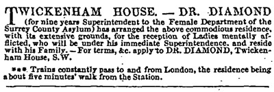
A few notes on Dr. Hugh W. Diamond and his home, Twickenham House :
Robert Wilfred Skeffington Lutwidge (1802-73). From an established county family in Cumberland. Educated St John's Cambridge. Barrister L.I. 1827. Auditor of the National Society for Promoting Religious Education 1828-43. Member of the Statistical Society from its foundation in 1834. Pioneer photographer and favourite uncle of Lewis Carroll. Lutwidge and Carroll shared their interest with Dr Diamond of Surrey County Asylum. Metropolitan Lunacy Commissioner 1842-45. Member of Commission of Enquiry into the State of Lunatic Asylums in Ireland 1856. Killed by a patient while visiting Fisherton House, Salisbury, Wiltshire. — The Anatomy of Madness, by W. F. Bynum, Roy Porter, Michael Shepherd (page 130).
IRONSIDES' HISTORY of TWICKENHAM. Miss LETITIA HAWKINS' ANECDOTES AND MEMOIRS. Wanted by Dr. Diamond, F.S.A., Twickenham House, Twickenham.
Marriages: Sept. 15. At Thurlby, Lincolnshire, Warren Hastings Diamond, M.D., eldest son of Dr. Diamond, F.S.A., of Twickenham-house, to Victoria Gonville, dau. of Sir Edmund Gonville Bromhead, bart., of Thurlby-hall, Lincolnshire. &msash; The Gentleman's Magazine (1863).
Seldom leaving his bed before noon, he breakfasted after a careful toilet, if a single cup of very strong green tea — drunk from the medium-sized breakfast-cup in the possession of Dr. Diamond of Twickenham House, Twickenham, — may be called a breakfast. He usually stood, whilst taking the tea, which was served for him without sugar or milk. — The Real Lord Byron: New Views of the Poet's Life By John Cordy Jeaffreson.
June 8.
Some notes on Dr. Henry G. Wright, a vice-president of the Photographic Society of London :
Dr. Wright,* of the Samaritan Hospital,
has recently called the attention of the profession to
the use of a compound of the iodide and nitrate of
silver as they exist in " an old photographic nitrate-
bath still bright and clear, but which had been so long
worked that it had become saturated with iodide of
silver, and contained a considerable amount of ether."
Accident led him to the use of this preparation, and
he has found it far more efficacious in the various
forms of stomatitis and analogous affections of the
uterus than the more concentrated solutions of the pure
nitrate of silver. Dr. Gibb has also used it topically
with marked benefit in affections of the throat and
larynx. This " old bath solution " may be obtained of
any respectable photographer. — Page 384, Clinical Notes on Uterine Surgery
by James Marion Sims (New York: Wood, 1886).
Footnote reference: [*]The Lancet, March 18, 1865, p. 282 :
" The Topical Use of Silver Solutions."
By Henry G. Wright, M.D.
In introducing the name of the newly elected Vice-President, the Council would refer to the number of photographers of eminence among the ranks of the medical profession. Many of them, like Dr. Wright, have been chiefly led to the study of the art through a recognition of its value in exactly delineating disease, without that irksome length of time required for a sketch even approximately accurate. The names of Dr. Diamond, of Dr. Arthur Farre, of Dr. Julius Dolfuss, of Dr. W. Budd of Bristol, of Professor Beale of Bang's College, deserve especial mention. And the beautiful microphotographs of Dr. Maddox and of Dr. Duchesne testify the importance of photography in demonstrating with unimpeachable accuracy the appearances of minute structure as revealed by the microscope. The time is probably not far distant when physiology will depend on photography for reliable illustrations of the subjecte on which it treats. — Page 191, Journal of the photographic society of London. (London: Taylor and Francis, 1865).
Dr. H. G. Wright sends in " A Portable Photographic Apparatus for Field or Room," containing some most excellent and portable arrangements, and which, with some different mode to give greater strength to the supporting legs, will be a valuable acquisition to the practical photographer. — Page 169, Journal of the photographic society of London. (London: Taylor and Francis, 1860).
The President then called the attention of the meeting to the very fine collection of Medical portraits and autographs now in possession of the Society, comprised— 1st, in the Soden collection, recently presented by Mr. J. Soden, of Bath ; and 2ndly, by the purchase of Mr. Squibb's collection, just sanctioned by the present general meeting ; both together forming a collection unrivalled by any in the kingdom. Notice was also taken of the rapidly-forming series of photographs, commenced by Dr. H. G. Wright, of pathological specimens, and the Fellows were urged to contribute to these collections as opportunities offered. — Page 298, Medical Times and Gazette. (London: Churchill, 1864).
It was stated in the Librarian's Report that 266 new works (independent of journals and continuations) had been added to the library, and, an increase of room for journals and serials being required, that shelving had been added, capable of containing 1800 additional volumes; also that a new feature had been added to the library, at the suggestion of one of the Fellows, Dr. H. G. Wright, in the shape of portfolios containing photographs of subjects of professional interest. A hope was expressed that Fellows possessing copies of such photographs would present them to the Society. — Page 279, Medical Times and Gazette. (London: Churchill, 1863).
On April 25th, 1863, the trifle laid before each diner's napkin was a small white cardboard diptych in a white paper wrapper. Containing on the one half of its interior a photograph of a familiar portrait of Shakespeare, and on the other half of the interior a photographic reproduction of Ben Jonson's no less familiar verses, beginning with ' This Figure that thou here seest put,' the diptych displayed on one of its outer surfaces nine musical and deftly- worded verses by Henry Wright, M.D. The paper- wrapper of the folded card displayed on the interior surface the following metrical note by Shirley Brooks :
As it fell upon a night,
Came a thought to H. G. Wright,
Loyal member of a club
That at Clunn's doth meet to grub. '
To its Shakespeare's feast,' quoth he, '
Something shall be sent by me,
And the Vanity is Fair
That could meet approval there,
Though I soar ceratis pennis
In the presence of Pendennis,
From the Second Folio (mine)
I will take the Portrait fine ;
Add the verses writ by Ben
On the foremost man of men ;
Get some rhymes (which here you see)
From the virtuous bard, S. B.,
And present the whole to each
Who shall hear our Thackeray's speech.'
SHIRLEY BROOKS.
Readers unfamiliar with the writings of the ' virtuous bard, S. B.,' may not regard as a fair example of his poetical address these rather feeble verses, which are interesting only because they relate to one of the latest banquets — possibly the last grand dinner at which Thackeray took the chair.
— Pages 306-307, A Book of Recollections. By John Cordy Jeaffreson (London: Hurst and Blackett, 1894).
A second work by Bitot — on the anatomy of the brain.
The more I study Charcot, the more I am convinced he is not understood by contemporary historians.
Began cataloging an important paper on contracture induced by hypnotism co-authored by Charcot and Richer.
Cataloged a paper by Caesar Boeck the great Norwegian dermatologist.
Began reading Bourneville and Pilliet.
Finished cataloging Babinski.
Began reading Babinski.
Paper on using photography to measure oxyhaemoglobin.
Today's link will bring up Voltolini's monograph on rhinoscopy.
Great, early, article on the medical uses of photography which describes doctors as pelicans.
So far as medicine is represented at the Centennial Exposition, much may be written. The Government has, in its own building and annexes, given the public an insight into the progress made in its medical departments. There is a building which is a model of an army hospital for twenty-four beds, built of the exact dimensions, and furnished with the same equipments as all other buildings of the same kind now constructed by the Medical Department. In this building models are exhibited of hospitals, hospital cars and vessels, as used in the late war. and the various contrivances which experience and ingenuity have suggested for the transportation of medical supplies, the sick and wounded, etc. One of the most interesting features here presented is the display of microphotographs, contained in portfolios, and placed in every window. Dr. Woodward, of the army, to whom the credit of such a display belongs, took an active part in the debate before a section of the American Medical Association at its recent meeting, maintaining the close identity of the blood corpuscles of man and some other animals when magnified, as some of these specimens, were 4350 diameters.
The library of the Surgeon-General's Office, or, as it has been recently re-christened, the National Medical Library, is represented here by a collection of albums of photographs, of uniform size, of title or specimen pages of more than a hundred curious books contained in it. One of these is a photograph of a page of a very old manuscript, Bernard de Gordon's " Lilium Medicum," bearing the date of 1349; another a photograph of the first printed monograph on diseases of children, by Paul Bagellardus, 1472 ; while the fifteenth and sixteenth centuries are largely represented in the list. Indeed, it seems like instituting a comparison at once between ancient and modern eras to find the bookmakers' productions of those distant days linked with the newer arts of the photographer, and thus vividly iutroduced to the thoughtful attention of others than mere bookworms or literary virtuosi. —The Medical Times and Gazette London: Churchill, August 26th 1876; page 237.
Transcribed from Google, an extract from Lionel S. Beale's book on the microscope, with reference to Delves. The book was published by Samuel Highley of London who also published Hugh W. Diamond's paper on photographing the insane. Here is the link to the Highley advertisement which appears at the front of Beale's book: Highley ad»»
PHOTOGRAPHY seems to he giving its powerful aid to medicine and its attendant sciences. Dr. Sanderson, in a paper on the influence of the heart examined by the movements of respiration on the circulation of the blood, gives a plan for registering the rapidity and volume of the human pulse, by means of the pulse- motion, which is made to record itself by a series of zig-zag lines on sensitivised paper. This may be considered rather a curious than a useful application of photography; but it is scarcely necessary to say that its aid is of the greatest value to the physiologist, the physician, and surgeon. The numerous changes made in the aspect of wounds can find a faithful record by no other means, and the splendid collection in the possession of the Royal Medico-Chirurgical Society is a testimony to the value placed by the profession upon this method of illustrating their science.
The power of the sun's pencil in giving minute and subtle indications of expression in the human face has made it a valuable teaching power in psychological medicine. The power of words to explain certain types of insanity is feeble as compared with the whole aspect of the patient and the expression of his face. These the photograph can give with unerring certainty. Dr. Conolly has illustrated a valuable series of papers on the varieties of insanity by photographs of the different types, taken by Dr. Diamond from his asylum, and as an aid to diagnosis they are truly valuable.
It is suggested that, before it is too late, the art should be made subservient to recording the types of the various races of men that are slowly disappearing as civilization advances. The physical aspect of man is a subject photography alone is capable of illustrating. — Edinburgh Review, No. 379-1371.
Began cataloging a second work by Recklinghausen — an album of photomicrographs.
Transcribed a paper by Dr. S. T. Stein on photographing the pulse with references to Marey, Vierordt, and Czermak.
Cataloged Sachs's landmark paper on congenital cerebral lipoidosis, also known as Sachs disease or Tay-Sachs.
Updated Forbes Winslow's psychological journal with a jpeg of the frontispiece.
Cataloged the eminent Jewish dermatologist, Heinrich Köbner. He established the first research facility for dermatology in Germany.
Finished cataloging Recklinghausen.
Began cataloging a monograph by the brilliant Recklinghausen.
Revised the Ferran description.
Finished work on the Ferran description.....for now and until the Spanish comes easier.
Began researching and cataloging Ferran's monograph on inoculation for cholera. It is an important document by a Spanish physician who had strong claims to the discovery of a vaccine against this disease and yet, there is no copy in the Surgeon General index and only 1 copy listed on worldcat. Strange.
Second paper by W. W. Keen, published in the Review. This completes the cataloging of all 48 articles that appeared in that journal.
Note to self :
PHOTOGRAPHS OF THE LARYNX. — We have had an opportunity of seeing Professor Czermak's photographs of the larynx and posterior nares. They are no less remarkable as giving perfectly distinct pictures of the glottis, the superior and inferior vocal cords and ventricles of Morgagni, than they are curious as the first and only photographs of the organ of voice. They give a striking proof of the advantages of sight gained by Professor Czermak's invention. On the 29th ult. Professor Czermak demonstrated the use of his instrument on several patients at the Great Northern Hospital. In one case the laryngoscope detected the existence of a constriction of the pharynx above the larynx, originating in cicatrisation and contraction of an old ulceration. This condition had not been previously diagnosed, as the patient, although labouring under partial loss of the soft palate and uvula, and suffering from complete aphonia and chronic sore- throat, had never complained of difficulty in deglutition. — THE MEDICAL TIMES AND GAZETTE, June 7, 1862, page 604.
Still another fatty tumor paper published in the Review. One more paper to go.
Finlayson claims that he took the first photograph of acromegaly in this paper.
Keen's plastic operation for protruding ears.
Interesting case of a gun shot wound to the head.
Another fatty tumor report in the Review.
Extroversion of the bladder by Maury.
Rupture of the bladder by Mears.
A third paper written by Samuel W. Gross and published in the "Review." He reports the surgical intervention in a case of traumatic aneurism of the carotid artery.
A paper by Dr. James R. Wood of Bellevue on a fatal case of elephantiasis. This document is important, not only because of the stature of Dr. Wood as the co-founder of Bellevue Hospital Medical College, but also because of the paucity of his published works.
A second paper by Andrews, published in the Review.
Scholar readers of this thread ought to check out Dr. Jill Bolte Taylor's talk on the two personalities of the human brain. It is the first inkling of what is becoming a transformative shift in how we structure and communicate information within our schools and institutions. The video is about 20 minutes in length and takes a few moments to load. The opening seconds are loud so if you use external speakers, turn down the sound. Here is the link : »»
Neuroanatomist Jill Bolte Taylor had an opportunity few brain scientists would wish for: One morning, she realized she was having a massive stroke. As it happened -- as she felt her brain functions slip away one by one, speech, movement, understanding -- she studied and remembered every moment. This is a powerful story about how our brains define us and connect us to the world and to one another. »»
A second paper by Duhring, published in the Review.
Encephaloid tumor of the hip capsule.
A second paper by Sayre, published in the Review.
Diverting from the Review to update the description of Barwell's monograph on orthopedic treatment of club-foot. Included four jpegs of the plates.
Beecher's second paper contributed to the Photographic review.
Updated Damon's monograph on leukemia. Included a jpeg of one of the plates.
Added an article by C. E. Webster on Applications of photography in surgery published in "The Photographic News." From that same journal comes these snippets:
Medical Photographs.—Dr. Richer and M. A. Londe have produced some remarkable medical photographs by the photo-electric apparatus previously described—the same subject having been reproduced successively at regular intervals, more or less frequent, at the will of the operator. — page 23.
PHOTOGRAPHY AS A MEANS OF OBTAINING PATHOLOGICAL RECORDS. — From the other side of the Atlantic we receive the following of the pen of Dr. A. L. Cory : - " As to the use of photographic outfits in medicine, I would say I find mine a great benefit. I have used it in cases of skin diseases, small-pox, spina befida, &c., and can see now where I should have kept photos of many cases if I had possessed it before. While in charge of Lake health department I took frequent copies of small-pox cases. It is so little trouble to keep the plate-holder filled and the camera in one corner of the consultation room. A photo of any case can be had at a minute's notice, the plate to be developed when convenient. I frequently take mine in the buggy when called to a case I think may be interesting, and use it if opportunity offers. Nothing that I know of offers us so easy and accurate a method of recording interesting cases." — page 399.
To the surprise of all, photography has quite failed in endeavoring to represent cutaneous diseases. It has been now pretty thoroughly tried, and the result is most unsatisfactory. We must agree with a shrewd observer and truthful recorder, Mr. Jonathan Hutchinson, that without color the photograph shows little or nothing of the disease ; and if hand coloring is put on, you at once destroy the special value of a photograph, its absolute accuracy as to detail. The test is this : Can you say what the disease is from the photograph without the name attached to it ? We have repeatedly failed. Moreover, to a large number of those published the real name of the disease photographed is not appended, but some other one. Hence photographs of cutaneous diseases have not been received with any great favor by professed dermatologists. As placards they, of course, serve a special purpose for both artist and doctor. — B. Joy Jeffries, Diseases of the Skin: The Recent Advances in Their Pathology and Treatment. Boston: Moore, 1871 ; page 76.
Townsend's second paper in the Photographic Review. This time his subject is an extraordinary hernia in a 40 year-old vagrant which began at infancy.
Small paper on hypertrophic diseases of the clitoris by an obscure Philadelphia physician.
A second paper by Samuel Weissell Gross, son of Samuel David Gross.
Extirpation of the thyroid. This was still a novel procedure in 1872, the first report for total thyroidectomy was written by Paul August Sick in 1867.
Paper on an adipose tumor over the sternocleidomastoideus.
Paper on genital warts.
Paper on aneurism in the abdominal aorta.
Another patient of Dr. Addinell Hewson with an epithelioma attacking the face. This time, however, the treatments with poltices of ferruginous clay were not successful and the patient died.
A short paper by S. D. Gross on deformity of the leg from a necrotic tibia.
This is perhaps the most important photograph published in the Review.
Dr. Maury, the physician photographer and editor of the Photographic review, lectured on the venereal diseases at Jefferson Medical College and helped establish the adjunct teaching hospital of that great institution. Today's link is his paper on a syphilitic gumma of the skull.
Elephantiasis græcorum, the title of this paper, is the antiquated term for leprosy.
As to the propriety of an operation in this case there can be but little doubt, as his general condition is excellent ; while as to the necessity, his comfort, happiness, and usefulness appear to be as much marred by this burden as was Bunyan's hero when he made his pilgrimage for relief.
Bladder polyps.
A misdiagnosis by a master!
Another photographic illustrated medical monograph by the physician photographer of Earth as a topical application in surgery.
A bicephalic monster which was on exhibition at Simpson's Museum and Menagerie in Philadelphia during the summer of 1871.
The science of American teratology began in Philadelphia with this paper by Pancoast on the pygopagus twins Millie and Chrissie Smith. The girls astonished the medical elite of Philadelphia with their quick intelligence and musical talent. The following quatrains were written by Christina:
'Tis not modest of one's self to speak;
But, daily scanned from head to feet,
I freely talk of everything,
Sometimes to persons wondering.
Some persons say I must be two,
The doctors say this is not true;
Some cry out humbug till they see,
When they say--great mystery!
Two heads, four arms, four feet,
All in one perfect body meet;
I am most wonderfully made
All scientific men have said.
None like me since the days of Eve--
None such, perhaps, will ever live--
If marvel to myself am I,
Why not to all who pass me by?
I'm happy, quite, because content,
For some wise purpose I was sent;
My maker knows what he has done,
Whether I'm created two or one.
The following poem, translated from the Hungarian, was written by an unknown physician
after his examination of the pygopagus twins, Helen and Judith:
Two sisters wonderful to behold, who have thus grown as one,
That naught their bodies can divide, no power beneath the sun.
The town of Szoenii gave them birth, hard by far-famed Komorn,
Which noble fort may all the arts of Turkish sultans scorn.
Lucina, woman's gentle friend, did Helen first receive ;
And Judith, when three hours had passed, her mother's womb did leave.
One urine passage serves for both ; — one anus, so they tell ;
The other parts their numbers keep, and serve their owners well.
Their parents poor did send them forth, the world to travel through,
That this great wonder of the age should not be hid from view.
The inner parts concealed do lie hid from our eyes, alas !
But all the body here you view erect in solid brass. — from: Gould and Pyle.
April 1.
When God plays the April fool:
V. Dr Byrom Bramwell showed (1.) PHOTOGRAPHS OF A CASE OF CYCLOPEAN MONSTROSITY. He met with it many years ago, when in general practice. The patient had borne five healthy children previously. Her sixth pregnancy terminated at the end of the seventh month, with twins. One child was naturally formed ; the other was the monster. The child was well formed otherwise. Dr Bramwell had unfortunately no opportunity of making a dissection. The child lived a quarter of an hour.
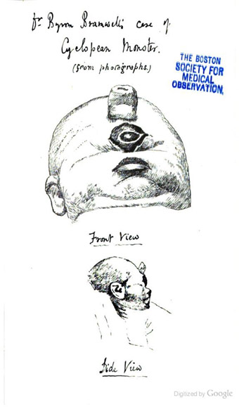
Began cataloging this paper by Sir Byrom Bramwell on intracranial aneurism. Bramwell was an accomplished artist and many of his texts are illustrated by drawings from his own hand:
The lithographs of naked-eye objects, represent with few exceptions the hearts of patients who have been under my own care during life, and with whose clinical histories I am intimately acquainted. The microscopical lithographs are, with two exceptions, copied from sections made by myself. In order to ensure absolute accuracy of representation, the naked- eye specimens were first photographed and then drawn under my immediate personal supervision, while the microscopical objects have been placed directly on the stone from my own drawings. — preface to Diseases of the Heart and Thoracic Aorta. New York: Appleton, 1884.
Believing that one great secret of all successful teaching is to teach by the eye as well as by the ear, I am in the habit of copiously illustrating my lectures by diagrams, drawings, and microscopical preparations. The diagrams and drawings are introduced into the text in the form of woodcuts, the microscopical sections are represented in colors. The chromo-lithographs are all drawn by myself, first with the camera lucida, and then in lithograph chalk ; they are with two exceptions ( figures 56 and 151, which are copied from Oharcot) representations of my own sections. — preface to, Diseases of the Spinal Cord. New York: Wood, 1886 (2nd ed.).
Von jeher pflege ich meine Vorlesungen reichlich durch Zeichnungen und Demonstration mikroskopischer Präparate zu erläutern, weil ich dafürhalte, eines der grossen Geheimnisse des erfolgreichen Unterrichtes liege darin, eben sowohl durch das Auge als durch das Ohr zu belehren. Diese Zeichnungen sind in der Gestalt von Holzschnitten in den Text des Buches aufgenommen; die mikroskopischen Präparate auf farbigen Tafeln wiedergegeben. Die Chromolithographien habe ich selbst — mit der Camera lucida und darauf mit der lithographischen Kreide — entworfen, mit Ausnahme der nach Charcot copirten Figuren 56 und 136, sind es Darstellungen meiner eigenen Präparate. — preface to German edition, Diseases of the Spinal Cord. Wien: Toeplitz & Deuticke, 1883.
Report on malformation of the fingers and toes by a prominent American surgeon who invented surgical instruments still in use today.
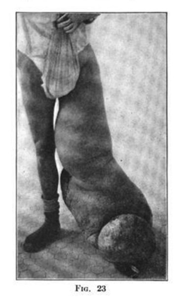
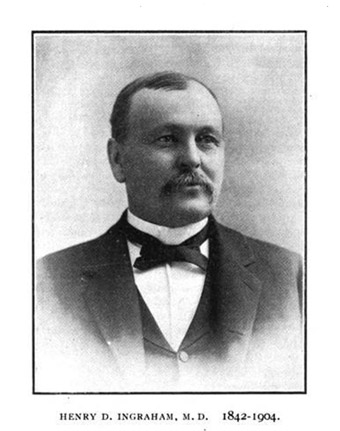
Todays' date is linked to a report on elephantiasis by Henry D. Ingraham of Buffalo whose obituary is posted below. Curiously, I found a similar image of elephantiasis reproduced on page 212 of the journal, Progressive medicine, (volume iv., 1907). The contributor's name is given as C[harles]. B. Ingraham of Johns Hopkins, but no relationship could be found with the Buffalo Ingrahams.
Dr. HENRY D. INGRAHAM died at his home in Buffalo, May 23, 1904, after a prolonged illness, aged 62 years. He was a native of New Hampshire, but his youth and early manhood were spent in the neighborhood of Arcade, .N. Y., where he was educated at the common schools and at the Arcade Seminary. For a time he taught in the common schools, and in 1863 began the study of medicine in the office of the late Dr. Lucius Peck, of Arcade. He attended medical lectures at the University of Buffalo, and graduated from that institution February 21, 1866.
Dr. Ingraham began the practice of medicine at East Randolph, N. Y., but soon settled at Kennedy, N. Y., where he associated himself with the late Dr. William Smith. He established there a large and lucrative practice, remaining until 1880, when he removed to Jamestown, N. Y., but after a few months decided to locate at Buffalo. He came here in 1881, and this city has since been the scene of his activities until his death. He developed a special liking for gynecological surgery and, while never entirely relinquishing family practice, he came to be recognised as one of the leading gynecologists, and in this department of practice he won the confidence and following of a large professional circle.
In 1883, Dr. Ingraham was one of the active organisers of the medical department of Niagara University, and until its union with the medical department of the University of Buffalo in 1898, he was its professor of gynecology and pediatrics. In the upbuilding of the department, he was an important factor. From 1898 to 1902, he was clinical professor of gynecology and diseases of children of the University of Buffalo. In 1883, he was appointed gynecologist to the Buffalo Hospital of the Sisters of Charity. Besides serving this institution most creditably as its gynecologist, he did much to advance its interests by supervising the erection of the new wing and other additions to the hospital, by cooperating with its authorities in many of the minor details of administration, and by aiding the establishment of the training school for nurses. After severing his connection with the Sisters' Hospital he became the gynecologist to the Riverside Hospital, continuing as such until his death. He was also one of the gynecologists to the Erie County Hospital from the time of its organisation until his death.
In medical societies he was active and influential. He was a member of the American Association of Obstetricians and Gynecologists, of the American Medical Association and also of the state, county and city organisations. In 1902, he was chairman of the section of gynecology and obstetrics of the Buffalo Academy of Medicine, and at the time of his death president of the Medical Union of Buffalo.
Dr. Ingraham was not a prolific writer, but he found time to publish occasional papers in the BUFFALO MEDICAL JOURNAL, the American Journal of Obstetrics and some other medical journals. No man of character and ability can live and work in a community for a quarter of a century without leaving his impress upon it, this being especially true of the successful physician, and such was Dr. Ingraham.
He is survived by a wife and step-daughter, by three sisters, and by several nieces and nephews, among the latter of whom is Dr. Henry C. Buswell of this city. A. A. H. — pp. 835-836 Buffalo Medical Journal, 1904.
Report from a surgeon who recruited and commanded in service of the Confederate army. His uniform can be found here »»
March 29.
BERLIN, Oct 3rd, 1890. The steady increase of the collateral branches, and the ever expanding utilization of physical forces, is gradually changing the character of medicine from a speculative to an exact and applied science. Photography, the latest handmaid of medicine, appears destined to play a vital ro1e in diagnosis, particularly in the zymotic diseases. Both in the Charité and the Clinicum it has been customary for some time to photograph patients with the view of thus securing typical aspects of affections, which might act as guides in future diagnosis. A few weeks ago a lady was admitted to the Charit6 for some uterine trouble and, as usual, was photographed. Imagine the surprise of the medical photographer when the portrait showed globular red spots disseminated all over the face. A picture taken on the following day presented a well defined eruption that after a week's time, became a clear case of small-pox. It is superfluous to dwell on the importance of the matter, and the only question presenting in this connection is whether it should not be obligatory, or if it is not at least desirable, to photograph every hospital patient at the time of admission. The Military Medical Academy will in future include photography in its curriculum of instruction. — page 490, The Medical Age: A Semi-monthly Journal of Medicine and Surgery, 1890.
Whoever visits Virchow of late is treated to the sight of a photograph of the "Hippocrates Tree," i. e., the tree under which Hippocrates delivered the first lecture on medicine. The growth stands in the market place of the Island of Kos, and as Hippocrates lived in 460-377 B.C., is 2,350 years old. Allow me to add here that the Father of Medicine died at Larissa, honored like a God, but poor as a church-mouse, for nearly all of his doctor bills remained unpaid. — page 491, The Medical Age: A Semi-monthly Journal of Medicine and Surgery, 1890.
Dr. W. F. Martin presented photographs illustrating the folio wing case: A boy 16 years, first came under treatment in June, 1890, suffering from alpecia areata, over a considerable space. — page 543, The Medical Age: A Semi-monthly Journal of Medicine and Surgery, 1890.
Here is an operation for soft tumor on the neck, reported by Joseph Pancoast, father of William H. Pancoast. Both father and son contributed to the Photographic Review. A woodcut of the photograph illustrating William Pancoast's first article is now posted and linked here: Horny tumors/ »»
March 28.
Dr Draper observed, that if a piece of metal, a shilling for example, or even a wafer, is laid upon a cool surface of glass or polished metal, and the glass or metal breathed upon, then, if the shilling is tossed from the surface, and the vapour dried up spontaneously, a spectral image of the shilling will be seen by breathing again upon the surface ; the vapour depositing itself in a different manner upon the part previously protected by the shilling.* More recently, Professor Draper has shown, that this spectral image could be revived during a period of several months of the cold weather in the winter of 1840-1 ; but he has stated that he cannot find the reason of this result, though he regards it as analogous to the deposition of mercurial vapour in the Daguerreotype. — Daguerre (1843), The Edinburgh review or critical journal: History and Practice of Photogenic Drawing, or the true Principles of the Daguerreotype, page 340.
[footnote] Dr. Keith has brought home with him from the Holy Land, about thirty Daguerreotypes of its most interesting scenery, executed by his son, Dr. George Keith, and which are now engraving for publication. Since this note was printed, we have received, and now have before us, fourteen of these beautifnl engravings, representing Mount Zion, Tyre, Petra, Hebron, Askelon, Gerash, Cesaræa, Ashdod, and other interesting places. — page 248, The Eclectic Magazine, 1847.
A paper on a child with inherited syphilis written by a physician who was so consumed by work, that he died at the age of 33.
March 27.
At the November meeting of the College of Physicians of Philadelphia, Dr. Weir Mitchell presented to the College some very interesting Harvey memorials which he collected during his recent visit to England and to the grave of Harvey, in the village church at Hempstead about seven miles from Saffron Walden in the county of Essex. In his visit to Hempstead Dr. Mitchell was accompanied by Dr. Benj. W. Richardson, of London, who has been principally instrumental in recalling to public notice the tomb of the great anatomist.
Dr. Richardson in early life (1847) was assistant to the surgeon at Saffron Walden, and while attending a neighbouring cottager's wife in her accouchement her husband entertained him with an interesting story of the chapel and vault of the "great Dr. Harvey." It gradually dawned upon Dr. Richardson that the Harvey referred to must be the discoverer of the circulation of blood, and on further inquiry this proved to be the fact. Up to that time the Harvey vault had not been visited by men of science within the memory of any of the surrounding inhabitants, and it had been long neglected and had fallen sadly out of repair. The villagers knew that Dr. Harvey was a celebrated man, who belonged to a distinguished county family, and had made some great discovery, but they did not know what it was. Dr. Richardson subsequently visited the vault and through his influence it was repaired and public attention called to the resting-place of the remains of the great Harvey.
In 1878, Dr. Richardson, with a photographer, made a visit to Harvey's tomb at Hempstead, and took a series of six photographs, copies of which Dr. Mitchell procured, had framed, and with fac-simile tracings of the inscription on the monument and on the sarcophagus of Harvey presented to the College. Two of these photographs represent different views of the exterior of the church, which dates back to the reign of Henry the Seventh, and one of its interior with the Harvey chapel, a handsome little annex over the Harvey family vault in the northeast corner of the church. Two others represent the front and profile view of the marble tablets or monument containing the bust of Harvey which is erected in the church. The ornamentation of the tablet is bold and effective, and below the bust is a lengthy and appropriate Latin inscription, a fac-simile copy of which Dr. Mitchell obtained by rubbing on tracing paper. A careful study of this bust which Dr. Richardson made in connection with Mr. Woolner, a sculptor and Royal Academician, led him to the belief that it was copied from a cast made after death, and that it was done with true artistic delineation, but that all that he was obliged to add to make up the bust as it stands, as for instance the ears, hair, drapery, etc., is of the worst possible quality.
The concluding photograph of the series is a view of the vault and of the sarcophagus containing the remains of Harvey which was obtained by the aid of the magnesium light. The leaden case or sarcophagus is represented as roughly shaped in the form of the body. The head has the rude, outline of a face with mouth, nose, and eyes ; the body is long and tapering towards the feet. The breast plate is broad and the inscription on it is in raised letters : " Docter William Harvey decesed the 3 of Ivne 1657, aged 79 years," a fac-simile copy of which Dr. Mitchell obtained by rubbing.
In accepting the gift of these interesting Harvey memorials the College voted its thanks to Dr. Mitchell.
— pp. 761-762, The Medical News and Abstract ; Philadelphia: Henry C. Lea, 1880.
Finished work on the paper by WW Keen and Thomson.
Began work on a paper by WW Keen and Thomson on a patient with gunshot wound to the head.
Report on Cholera in Australia illustrated by the author's own photographs.
Paper on a segmented urethral calculus removed by T. G. Morton, the chief surgeon of Pennsylvania Hospital and husband of Kirkbride's daughter.
March 10.
She then came again under his care, with an enormous waxy liver, and in a hopeless condition. The limb was slightly flexed and adducted, and one sinus existed over the left hip, from which pus escaped. She suffered no pain, but remained quietly in bed, the urine and faeces were passed involuntarily. She lingered on for four months, when it was noticed that there was a very sharp prominence over the spinous processes in the dorsal region, and one also in the lumbar region. The breathing became rapid and laboured ; and steadily sinking, the child died three years and six months after the operation.
On post-mortem examination, the organs were, as was diagnosed during life, in a waxy condition, and traces of old tubercular disease were found in the lungs.
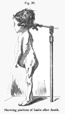
After death, a photograph, from which the accompanying woodcut was obtained, was taken by Mr. Mason, of Bellevue Hospital, by simply suspending her in a head rest. It will be observed that the limbs are nearly normal in position, and they assumed this position by their own gravity, without any extension or traction being applied to them. The limb operated upon is, in fact, the straighter of the two, and is not so much flexed at either hip or knee as the other. A sharp angular projection is distinct over the third dorsal, and another not so prominent over the first lumbar, vertebra; the enormous abdomen is markedly conspicuous.
Length of body, 30 inches ; left lower extremity 13 inches from anterior superior spine of ilium to external malleolus, right limb 13J inches long between same points. Length of both limbs from trochanter major to external malleolus 13s inches (these measurements were made by Professor Stephen Smith). Showing position of limbs after death. — p. 337, Transactions of the International Medical Congress, Seventh Session, Held in London London: J. W. Kolckmann, v. 2, 1881.
A remarkable recovery from excision of the hip joint.
March 9.
The Finsen Institute is located amid the shady boughs of great trees in the edge of the town. About it are numerous private villas. Rovsing's clinic is nearby. The institute consists of only two buildings; one, the laboratory, is an old villa. The clinic building was especially built for Finsen's work. At the left, as one enters the grounds, is a little low red building that one does not notice until attention is called to it. This was the place where Finsen first worked out his ideas. The building was brought here from another part of the city, and serves as a memento of the beginning of Finsen's efforts. If you enter the clinic suddenly you are somewhat startled at first. There are perhaps half a hundred patients in the big room, waiting their turn at the light machines.
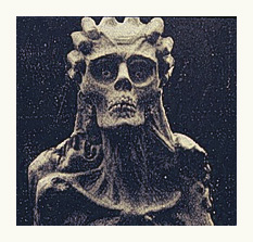
The faces you encounter make you shudder. It is like a first view of Boleslas Biega's sculpture. Even the white, expressionless, cicatricial faces of the cured cases one has to get used to. But the horrible disfigurement of advanced, untreated lupus vulgaris is terrible. One face was a blank, reddish-white mass, ringed with two pink circles, from which dull eyes glanced staringly; there was no nose, and a ragged hole with everted, granular border, served for mouth. No wonder they honor the name of Finsen, when he has given to his people the means whereby so hideous a human being can be restored to a fair semblance of his original self.
The patients, many of whom have come from distant parts of the globe, are first photographed and then seen by a physician, who rings, with a wax pencil, the exact spot to which the light is to be applied. Then they are taken to the operating-room for treatment, after which a simple ointment and a bandage are applied. That is all. Some cases need only a few treatments, others must remain for many weeks. Patients are advised to come back in six months or a year to have some spots that may have escaped the rays cleaned up. Each treatment costs from fifty cents to a dollar according to the circumstances of the patient. — pp. 34-36, Glimpses of Medical Europe by Ralph Leroy Thompson (Philadelphia and London: Lippincott, 1908).
A paper on what is probably a neurofibroma of the face.
Work on the bibliography resumes with this paper by John Hill Brinton, a doctor who in his role as the first director of the Army Medical Museum, made significant contributions to medical photography.
Paper by S. Weissell Gross, son of S. David, on cystomatous tumor of the perineum in a male subject.
This paper on fibroma in the clitoris is interesting for Bumstead's comment on adventitious growths.
Skipped to volume 2 of the Photographic review of medicine & surgery for this paper on vascular tumor of the lips, by an anatomist at Jefferson Medical College.
Maury's case of a former slave suffering from keloids that developed after severe physical abuse and trauma. It is an American story.
Pancoast's famous case of exuberant horny excrescences on the face of a barnacled old sea captain.
Samuel David Gross led the inaugural issue of the Photographic review of medicine & surgery with this article on echinococcal cyst of the thigh.
Bibliographical work begins anew with cataloguing the individual contributions in the journal, Photographic review of medicine & surgery.
A letter by Galton on composite photography which was published in The Photographic News (1888).
January 28.
Theodore Schwann, the author of the cell-theory. To some of our readers it will be a startling piece of intelligence that the founder of modern histology is actually at this moment alive, and teaching as Professor of Physiology in the Belgian University. The committee charged with the management of the celebration desire the co-operation of scientific bodies and of individuals in this country. We are authorised to draw the attention of officials of the learned societies and other corporations to the approaching event, and to beg them to obtain some expression of sympathy with the object of the celebration—viz., the doing homage to the genius of Theodore Schwann. It is requested that letters intended to be read at the celebration may be forwarded either direct to the secretary, Prof. Edouard van Beneden, Liege, or to Mr. Ray Lankester, Exeter College, Oxford. All Englishmen of science who have specially occupied themselves in the field of work opened up by Schwann, are begged to communicate individually with either of the above- named gentlemen, and to forward their photographs for insertion in an album which is to be presented to the founder of the cell-theory. — from the journal, Nature, March 28, 1878; page 436.
Swann auctions is offering an original albumen print from the Duchenne masterpiece, Mécanisme de la physionomie humaine/ (Figure 8) at its sale on February 7, lot #47. The first 109 lots, which include the Duchenne and another medical subject (X-ray of the hand... lot 48), were placed by "An American collector" who has written a prefatory paean on his experiences in the hunt of photographic stories. His essay is a superb read and I recommend it to scholars of all genres of photography.
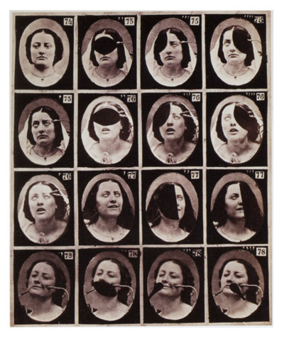
Transcribed from Galton's autobiography, his chapter on composite photography. Also have had the pleasure of corresponding today with Dr. Laurie Slater, a physician and scholar of medical antiquities who is generous in sharing his knowledge through his website, Phisick. Mahomed's sphygmograph with ivory rests is one of the treasures displayed there and can be found under the "Medicine" category on his menu. The brilliant Dr. Mahomed collaborated with Galton on his phthisic physiognomy essay which was catalogued yesterday and in the chapter of Galton's autobiography transcribed today there is this following anecdote:
With the help of Dr. Mahomed and the permission of the authorities of Guy's Hospital, I took many photographs of consumptive patients and made composites of them, which are published in the Guy's Hospital Reports, vol. xxv. They show two contrasted types, the one fine and attenuated, the other coarse and blunted. Dr. Mahomed was a very promising physician, on the eve of becoming well known, when he caught a fever of the same description, I am told, as that on which he had become an authority, and died of it in his newly purchased house.
Finished Roux. Began cataloging another article by Galton, illustrated with his composite photographs of phthisic physiognomy.
I will conclude with a simile. The final verdict about works of art and men of genius may be compared to one of those composite photographs (devised by Mr. Francis Galton which are obtained by the superposition, one above the other, of many negatives taken from different individuals. Each separate face has left its filmy impress on the composite photograph; and all the faces have contributed to form a type the type of a criminal, the type of a consumptive person, the type of a certain family. Blurred in some of its outlines and details as the ultimate result may be, such a composite photograph has an unmistakable generic individuality, which is even more instructive, even more convincing for the student of criminal, consumptive subject, specific family, than the mere aggregate of single photographs which compose it. It yields, not the person, but the type. Even so the final verdict of criticism is the total result of countless personal judgments, superimposed, the one above the other, coalescing in their points of agreement, shading off into blurred outlines at points of disagreement, but combining to produce a type which is an image of fundamental truth. — "The Criterion of Art," extract, from Essays Speculative and Suggestive, (1890) by John Addington Symonds.

The individual photographs were taken with hardly any selection from among the boys in the Jews' Free School, Bell Lane. They were the children of poor parents. As I drove to the school through the adjacent Jewish quarter, the expression of the people that most struck me was their cold, scanning gaze, and this was equally characteristic of the schoolboys. The composites were made with a camera that had numerous adjustments for varying the position and scale of the individual portraits with reference to fixed fiducial lines; but, beautiful as those adjustments are, if I were to begin entirely afresh, I should discard them, and should proceed in quite a different way. This cannot be described intelligibly and at the same time briefly, but it is explained with sufficient fulness in the Photographic News, 1885, p. 244. — extract, "On the racial characteristics of modern Jews," by Joseph Jacobs, in: The Journal of the Anthropological Institute of Great Britain and Ireland, (1886).
Finished transcribing Duchenne's essay. Began cataloguing a Roux treatise on amputation at the ankle.
Began transcribing Duchenne's essay titled, Recherches icono-photographiques sur la morphologie et sur la structure intime du bulbe humain.
A monograph on surgery of the infant palate by a master of the art of uranoplasty. Ehrmann presents 41 operations for repair of the cleft palate. The book is illustrated by 62 photos of casts made before and after the operation, however this information is unverified until I find a copy.
Finished cataloging the Bonnet biography. Scientia has a signed presentation copy of Bonnet's major opus, Traité des sections tendineuses et musculaires/ and Norman has Bonnet's grand opus, Traite des maladies des articulations (with the atlas). Both works are treasures that will be picked up by a savvy collector.
January 18.
Began researching a little biography on the French orthopedic surgeon, Amédée B. Bonnet (1802-1858). Here is a passage about Bonnet written by Leonard F. Peltier and transcribed from page 36 of his book, Orthopedics: A History and Iconography (Norman Publishing, 1993):
In Lyon, the development of the new specialty was in the hands of Amédée B. Bonnet (1802-1858) (figure 2.23). Bonnet studied in Paris in the early part of the eighteenth century, when it was the center of the medical world. Receiving his medical degree in 1832, he went immediately to Lyon, where he became associated with the Hôtel Dieu. His practice flourished and he eventually was made the chief surgeon of the hospital and professor of surgery in the medical school. While he wrote on many surgical subjects, it is his work on joint diseases that remains most important. He believed in treating his patients by operative methods as well as by nonoperative means such as manipulation, splinting, and immobilization. His book, Traité des sections tendoneuses et musculaires, which appeared in 1841, discusses the use of subcutaneous tenotomy in the treatment of strabismus, myopia, stammering, clubfoot, deformities of the knee, torticollis, fractures, and other conditions.
Italian psychiatrists.
Here is an 1883 Marine Hospital report on fracture of the skull at the base. Illustrated by two large photo plates.
Work done today on describing Aeby's monograph on the lung.
Added more information pertinent to the Ewart monograph.
Above is a link to the essay on gelatine-bromide emulsion which revolutionized photography.
Below is a description of marking tales, hand-written into a book listed on eBay »»
The doctor writes that if a woman who is pregnant gets scared or stressed, her baby will be affected and he gives three case examples. A women who encounters a beggar on the street crawling to her with deformed legs-she then gives birth to a baby with deformed legs, another woman's husband shots and kills a squirrel or was it a cat, and shots its head off, she gives birth to a headless baby, another who's husbands slaughters cows ask for her help, her baby at a young age of childhood goes off to slighter woman.... hello! this is 1880s and the doctors think that!
Here is Ewart on the morphology of the lung, illustrated with photogravures.
Spent a little more time researching FS Watson and adding to the description. Began researching Ewart.
Finished the description for the Watson atlas.
First entry of the year is this monograph/atlas by Francis Sedgwick Watson on the diseased prostate, published in 1888. This is the personal copy of Herbert Leslie Burrell, (1856-1910) and came with two envelopes of illustrations from Watson inserted among the pages. Possibly Burrell meant to use them for a surgical compendium he was planning, but never completed. Photographs will be posted tomorrow.
