Journal : Photographic review of medicine & surgery ; vol. 1., no. 1.
Philadelphia : J. B. Lippincott & Co., 1870-71.
Description : pp. 8-10, [1] pl. ; ill.: 1 photo. ; 24 cm.
Photograph : mounted albumen.
Subject : Skin — Cicatricial keloid.
Notes :
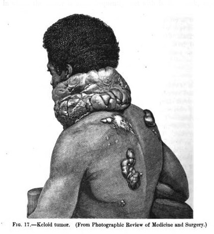
Dr. Maury's photograph of a quondam slave with extensive keloids also appeared as a wood engraving on page 668 of John Vietch Shoemaker's dermatology textbook, titled, A Practical Treatise on Diseases of the Skin (New York: D. Appleton, 1901):
FREDERICK JOURDAN, colored (formerly a slave), aet. 28 years, admitted to the surgical wards of the Philadelphia Hospital, May 26th, 1870. Is a native of North Carolina; he is above the ordinary height, of large frame, and very muscular. His family history revealed nothing from which to trace the cause of his disease. Thinks his father had two small tumors, one under the chin and the other on his side, both of which gradually disappeared before his death, the cause of which he does not know. Mother and brothers alive and in good health.
When eight years of age a small abscess made its appearance on the anterior part of the neck, which, on being opened, discharged an ounce of pus. As the result of this abscess a well-marked induration followed at the original seat, which gradually extended in both directions around the neck. After nine years' growth it had half encircled the neck, and was about two inches in width. Professor N. R. Smith, of Baltimore, at that time removed the growth. The resulting wound healed kindly in six weeks. The line of the cicatrix, however, was speedily occupied by a hard, rounded ridge, which slowly extended and enlarged. Eighteen months later, an accidental wound was inflicted by an axe on the posterior part of the neck. This wound also soon presented a hard, nodulated cicatrix, which crept around the neck to join the one from in front.
His master now carefully avoided punishment, as the least incisions seemed prone to take on this morbid action. Four years after this date a band of soldiers whipped him severely ; each gash on healing was succeeded by the hard, elevated ridge, the result of the previous wounds.
Seven years subsequent to Dr. Smith's operation, the tumor was again removed, at which time it had extended around the entire neck. Three months were occupied in the healing of the wound, which also assumed the same morbid action.
At the present time there are thirty-seven tumors of variable size. The large one resembling, in a marked degree, the ruffles worn in the time of Queen Elizabeth. The two original growths are now thoroughly blended and form one solid mass, touching at the posterior part of the neck. It measures twenty-eight inches in its greatest circumference, and five inches in its perpendicular diameter. It is plicated, and has deep fissures separating the folds, from the bottom of which is exuded a thin, yellowish, offensive fluid. The skin is intact, not having undergone ulceration. There is little or no elevation of temperature, no pain when pressure is instituted, it being only painful from the weight. The entire mass can be moved without difficulty, thereby indicating only a cutaneous attachment. The sensibility of the skin is perfect, the presence of a fly being at once recognized.
The other tumors are situated on the back and right arm, and vary from the size of a pea to that of a medium-sized tomato.
In July, 1870, two of these growths were removed for the purpose of microscopical examination, and also to observe the rapidity of recurrence. For one the ecraseur was used, for the other the knife; in both instances a portion of healthy skin tissue was removed. At this date, almost three months afterward, there is a well-marked tendency to the development of new growths in the same place.
The following careful microscopical examination was kindly made by Dr. L. A. Duhring :
"To the naked eye, upon section vertically through the tumor, the cut surface presented a structure close and compact in appearance, of a yellowish-white color. To the touch it was tough, resisting, and firm, with a certain amount of elasticity, and upon pressure exuded a thin, pale-straw colored liquid."
"After being prepared in solution of bichromate of potassa and alcohol, vertical sections were made and examined in glycerine. The horny layer of the epidermis was thin and scanty, the cells themselves being well broken up, and many of them having undergone granular degeneration. The cells in the upper layer of the rete mucosum seemed closely packed together and unusually numerous, while the deeper layer contained the pigment cells well colored.
"The mass of the tumor was composed principally of connective and elastic tissues, the former being disseminated throughout, while the latter appeared here and there in the form of good- sized, well-developed elastic bands, running both transversely and vertically. Fat was found in some parts in fine globules. Long, wavy bundles of connective tissue were seen running in striae transversely, just beneath the papillary layer. Here and there a cut sebaceous gland was found.
"In some of the fields a loose network of connective and elastic tissue intermingled, was present, with globules of fat. Connective tissue cells, long and twisted, were to be seen, sometimes approximating each other and again scattered."
As respects the nature of this affection little doubt exists in my mind. Although not strictly corresponding to the keloid of Alibert, nevertheless its history, physical appearances, and microscopical nature undoubtedly place it with that group of tumors that modern pathologists denominate keloid. Professor Gross, in his work on Surgery, vol. i. p. 583, describes this affection.
The error into which one would readily fall exists in the name, inasmuch as a keloid formation is generally accepted to be a tumor crab-like in appearance, and resulting frequently from operative interference with other tumors; but at the same time a " keloid diathesis," as it were, may exist, where the least incision into the skin is surely followed by this abnormal condition, then the growth does not present the peculiar appearance which has procured for it this name. As for example, in the present instance, each gash of the lash has been replaced by a tumor resembling a tomato in shape and pediculated, yet careful microscopical research reveals nothing except a fibro- plastic element common to other formations. Whereas the history of the case, the physical condition, and collateral evidence will do much to establish correct facts.