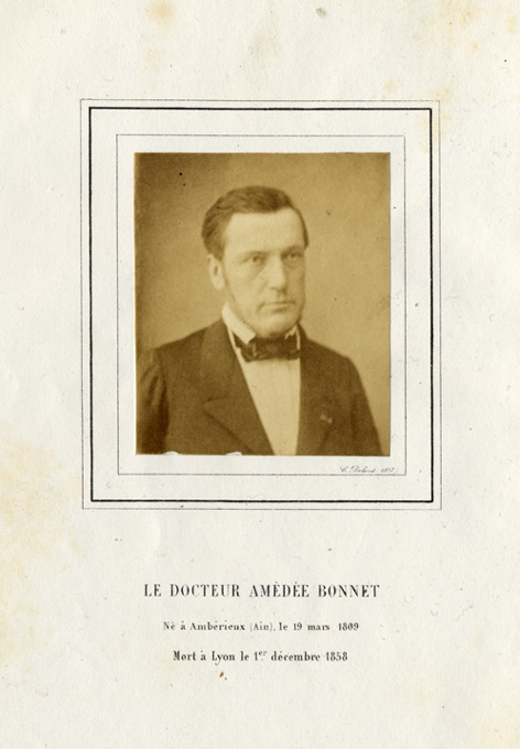
Avec un portrait photographié par Dolard.
Lyon : Aimé Vingtrinier, 1859.
Description : [i-viii], [1]-170 p. ; ill.: 1 photo. front. ; 25 cm.
Photographs : 1 frontis leaf with mounted albumen.
Photographer : Camille Dolard (Dolard jeune), 1810-1884.
Subject : Medical biography — Amédée Bonnet.
Notes :
Toutefois, le grand ouvrage de cette époque de la vie du professeur Bonnet fut son Traité des sections tendineuses et musculaires (1841). La méthode des opérations pratiquées sous la peau, c'est-à-dire à l'abri du contact de l'air, venait de naitre. A. Bonnet se précipite dans la mêlée un peu confuse des inventeurs et y prend, dès l'abord, la première place. Son livre, plein d'aperçus neufs et profonds, éclaire d'une lumière inattendue l'anatomie des enveloppes de l'œil, le traitement physiologique de la myopie et du bégaiement, la théorie du pied-bot et des déviations du rachis. Ce fut là le premier et solide échelon de sa grande renommée: des opérations multipliées de strabisme et de pied-bot rendirent son nom populaire; son ouvrage lui donna dans la science une position qui ne pouvait plus que grandir. D'ailleurs, ses travaux sur les Positions des membres dans les maladies articulaires et sur les Appareils de mouvement étaient déjà comme des jalons de l'œuvre capitale de sa vie, de son Traité des maladies articulaires, qui devait lui faire un nom européen, en lui créant un rang à part dans la chirurgie moderne. — Garin (page 5).
One important feature of tuberculosis of the peripheral joints was the consistency of the deformities produced. Each joint tended to assume a characteristic posture as it was affected by the disease. The explanation of this phenomenon was supplied in 1840 by Amédée Bonnet (1802-1858)[sic] of Lyon, who carried out an extensive series of experiments on fresh cadavers of all ages. By injecting fluids under pressure into the capsules of the joints, he observed that the limbs assumed the positions that permitted the greatest amount of fluid to be injected. He also noted the points of rupture, or the weak spots, in the capsule. He correlated all of his findings with clinical observations on his patients. The hip assumed the position of flexion, adduction, and external rotation; the knee, flexion, posterior subluxation, and external rotation of the tibia (figures 6.30-6.31). This beautifully carried out investigation is well worth reading today as an example of the scientific standard that was achieved in the first half of the nineteenth century. — Leonard F. Peltier (Orthopedics: A History and Iconography. San Francisco: Norman Publishing, 1993; pp. 36-37).

Bonnet was probably the first to recognize the sanguinous effusion and anchylosis which commute from extended immobilization of a joint. His reputation for subcutaneous tenotomy transcended borders, advancing this procedure in the treatment of many conditions including strabismus, myopia, stammering, clubfoot, deformities of the knee, and torticollis. (1841) He was the first to operate, successfully, for the extraction of intra-articular disease bodies and in ophthalmic surgery he established the standard for enucleation of the bulb, still in use today. (1842)
The photograph reproduces with cropping an original photograph now in the archives of the Académie nationale de médecine»»
