Journal : American Journal of Otology ; vol. iv.
Boston : Houghton, Mifflin & Co., 1882.
Description : p. 186, [1 l.] pl.; ill.: 1 photomicro. ; 24 cm.
Photograph : 1 photolithograph.
Photographer : John W. S. Arnold, M. D., Emeritus Professor of Physiology and Histology, New York University College of Medicine.
Subject : Ear — Tumors.
Notes :
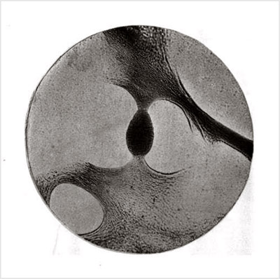
The accompanying plate is a photolithographic copy of a micro-photograph taken by Professor J. W. S. Arnold, M. D., of this city. It represents one of those minute pear-shaped bodies which Professor Politzer and Professor Kessel discovered, independently of each other, in 1869. (See Vols. V. and VI. of the Archiv fur Ohrenheilkunde.) In Professor Politzer's communication two rather coarse wood-cut illustrations of these structures are given, but, so far as I can learn, these are the only cuts that have as yet been published. The specimen here represented is so perfect in all its connections, and the photographer's work has been so well done, that it seemed to me that a photolithographic copy was worthy of being preserved in a journal,devoted to otological matters.
So far as the specimen is concerned, from which the photograph was taken, I may say that it was prepared in the autumn of 1869, in the laboratory of Professor Julius Arnold, in Heidelberg. It represents a horizontal section through the stirrup and surrounding bony structures of the oval window. The membranous net-work shown in the plate was found stretched out between the posterior limb of the stirrup, the tendon of the stapedius muscle, and the wall of the niche for the oval window. The fresh specimen — which, if I remember rightly, was obtained from the body of an infant, — had been treated first with a very weak solution of chromic acid and hydrochloric acid, and then with absolute alcohol. The section itself was treated with oil of cloves and mounted in damar varnish. The photograph was taken directly from the slide by Professor J. W. S. Arnold, in the early part of 1870, and the magnifying power of the lens employed was about fifty diameters.