Journal : Photographic review of medicine & surgery ; vol. 2.
Philadelphia : J. B. Lippincott & Co., 1871-72.
Description : pp. 15-16, [1] pl. ; ill.: 1 photo. ; 24 cm.
Photograph : mounted albumen.
Subject : Lips — Vascular tumors.
Notes :
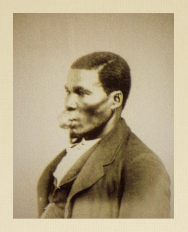
SAMUEL. PINKER, aged 19, a negro and a native of Delaware, presented himself at the clinic of the Jefferson Medical College. The history which he gave was as follows : When about ten years of age he received a blow with a stick upon the upper lip, producing a slight incised wound of the surface. The contiguous part immediately swelled and active inflammation ensued, which in time subsided, leaving, however, a permanently enlarged lip. This swelling gradually increased in size until, six years having elapsed since the occurrence of the accident, it attained the size of a walnut. Though not suffering any inconvenience from it at the time, he feared the development of malignant disease, and sought the aid of a surgeon who advised and performed an operation, the nature of which was unknown to the patient.
The operation, however, had not the desired effect and the tumor increased in size until it assumed its present dimensions. The photograph represents him three years after the so-called operation, as he presented himself at the clinic. Upon examination we found a large tumor of the upper lip, the size of a hen's egg.
Almost its entire surface was mucous membrane, although from exposure it had assumed the character of skin. In consistence it was soft and pliable, its bulk diminishing under pressure, but regaining its size when this was removed. There was no pulsation nor increase of temperature. His general health was good.
Many plans of operation have been suggested for this class of tumors, excision and the injection of various liquids being the most common.
The one deemed most desirable by Prof. Pancoast in this case was that of excision. To control any excess of hemorrhage which might possibly occur, the base of the tumor was transfixed by means of a large acupressure pin, which was passed through the external surface in a transverse direction. An incision was now made along the mucous surface of the lip, and parallel with it, the mucous membrane being dissected up, and the mass of distended capillaries removed. This was found to be quite small, the loss of blood having diminished the bulk in an extraordinary degree. The wound was allowed to remain open until all hemorrhage had ceased, which, however, was slight. The edges were then brought together by the ordinary interrupted suture, and afterward cold water dressing applied. On the third day the pin was removed, the patient progressing favorably. On the fifth day the stitches were removed, and five days later the man was permitted to return to his home. Upon examining the mass, I found that it coincided precisely with the description given by Prof. Gross in his System of Surgery, vol. i. p. 849 : " The venous tumor consists of a network of dilated capillaries, not of new development, but simply an exaggeration of those which are peculiar to the part where the morbid growth is situated. They are connected together by a loose cellular tissue, are more or less tortuous in their disposition, and have exceedingly thin, delicate walls, so that when a body of this kind is excised, they immediately collapse, the structure which previously formed a large mass sinking away into a little, spongy remnant."
Thomas Hollingsworth Andrews served as Demonstrator of Anatomy at Jefferson Medical College from 1875 to 1879 under Professor William Henry Pancoast.