Journal : Photographic review of medicine & surgery ; vol. 2, no. 4.
Philadelphia : J. B. Lippincott & Co., 1871-72.
Description : pp. [27]-29, [1] pl. ; ill.: photo. ; 24 cm.
Photograph : mounted albumen.
Subject : Hip joint — Anchylosis.
Notes :
C. R., aet. 24 ; single, native of New York ; teamster. Admitted into Bellevue Hospital January 4, 1872, with the following history :
About the middle of January, 1871, while attempting to lift a barrel of nails into the back of his wagon, he felt something give way low down on the inside of his back, and at the same time a severe pain in the inside of both hips and groins, but most severe on the left side. This was followed in a few weeks by a bubo or swelling in each groin, and as he had a slight urethral discharge at this time, it was suspected to be sympathetic with this difficulty, as no mention was made to his then attending physician of the previous muscular strain. He was sent to the Strangers' Hospital March 10, 1871, and I am indebted to my friend, Dr. F. S. Otis, one of the physicians of the above hospital, for the following notes, copied from the case-book, headed " Abscess in Track of Vas Deferens."
"On admission, patient a strong, healthy man. In both groins decided induration exists ; slight fluctuation on left side, with tension.
"March 12th.—Abscess in left groin opened; very little pus, and some blood, discharged.
"March 13th.—Opening enlarged to prevent burrowing; bubo stuffed with cotton.
"March 15th.—Tenderness in scrotum of left side, with hard swelling, extending from external abdominal ring to the outer side of the vas deferens and just over the left crus of the penis; very painful to the touch, but giving no impulse on coughing, and slightly movable. . . . .
"March 31st.—Explorative operation performed by Dr. Otis; Drs. Bumstead, Sands, and Sabine present. A straight incision was made through the scrotum on the left side, and the mass fairly exposed. It was found to be closely connected with a hernia above, from which it was detached with the scalpel. The mass was hard, and at the same time very friable, the finger penetrating it without much force, and on so doing a little pus escaped. A piece of the mass an inch long was removed for examination,* and the wound stuffed with lint." . . . .
The daily record of the case is very interesting, but too extended for this paper. I can only sum it up by saying, that he had excessive suppuration, hectic fever, and great prostration, followed in a few weeks by severe muscular contraction; and on April 28th the notes state :
"The thigh is drawn up at a right angle to the body. He is unable to relieve it. Motion in knee perfect. . . . .
"Extension was applied at various times with different weights, but could not be borne on account of the pain produced. The notes state :
"June 1st.—Sinus has healed. . . . His condition is pitiful ; . . . unable to extend the left thigh and leg, which is still bent at an angle of 100° with the body, and also adducted, so that the knee points out to the right side." . . . An extensive slough formed over the left trochanter major, owing to the extreme pressure of it against the soft parts, from the strong adduction of the thigh.
"October 17th.—Sinus has finally healed. Patient is as strong as ever. There is great deformity of the left lower extremity. Whole pelvis is oblique, left side being the highest; the thigh is still flexed, but not so much as previously, and is drawn over to the opposite side. There is tonic contraction of the adductors, flexors, and hamstring muscles; most marked in the former. Discharged."
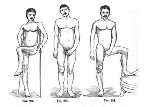
When he presented himself at Bellevue Hospital he was carefully drawn by Dr. Leroy M. Yale, from which the photograph (Fig. 1 ) was taken.
His limb could be drawn nearly parallel with the other, but it was done by rotating the entire pelvis on the opposite acetabulum, and raising the crest of the left ilium nearly four inches higher than the opposite side.
January 10, 1872—I operated in the amphitheatre of Bellevue, in the presence of a large class, and a number of physicians of the city, among them Drs. J. C. Nott, McIlvaine, Henry, and others. My house-surgeon, Dr. Cushing, had previously fitted to the right side of the patient's body a plaster-of-Paris mould, extending from the axilla to the foot, for the purpose of counter-extension, when the abduction should be applied after the operation. Chloroform having been administered, I divided the gracilis and the adductors subcutaneously, closed the wound with adhesive plaster, and applied a figure-of-eight roller. Then laying him on his back, and placing my knees on either ilium to hold his pelvis, I forcibly broke up the remaining adhesions, and succeeded in bringing the limb into position. Adhesive plaster, for extension, was secured to the whole limb by a roller, and the plaster-of- Paris mould secured to the right side of the body and limb by another roller.
The patient was then placed in bed, and extension and abduction kept up by weight and pulley. Ice-bags were applied around the hip. The wound healed without any suppuration, and no unpleasant symptoms followed the operation.
February 22, 1872.—Patient walked from my office to the photographer's, and had Fig. 2 taken, which shows his complete recovery.
* Under the microscope nothing malignant was found.
After making slight modifications, Sayre republished this paper in his monograph, Lectures on orthopedic surgery and diseases of the joints. The photographs were reproduced as woodcuts in that work.