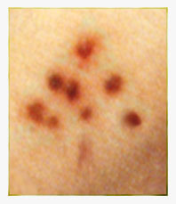

Just found this informative article on Duchenne that was written by André Parent and published in the Canadian Journal of Neurological Sciences this past August. Title of the article is, Duchenne De Boulogne: A Pioneer in Neurology and Medical Photography. If the links here do not bring up the PDF file, go to http://cjns.metapress.com and on the search page enter "Duchenne" in the title field and "Parent" in the author field and it should come up. The article provides information about an unpublished atlas by Duchenne comprised of the first photomicrographs of brain neurons and nuclei and I hope to enter it in the Cabinet bibliography when I can find its location.
More work accomplished on the index of nerve disorders.
As promised, here is the rough draft of the index of nerve disorders. One or two of the subjects will be moved to another index, but the list should be double in size when it is done.
My recent work is somewhat random. Added more images to the index of skin disorders and began work on the neurological disorders index. Sorted through slides. Did more work on the Dr. Weeks portfolio.
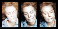
My attention was drawn to these three slides of traumatic shock syndrome and I couldn't resist the diversion of this beautiful triptych :
Index of skin disorders is now pretty much complete. Next project will be an image index for neurological disorders.
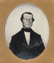
For the opening page of the Clinical Portrait menu, I am using a detail from this ambrotype of a man probably suffering from Bernard-Horner Syndrome. Several of the signs are there : a partial ptosis of the right eyelid, enophthalmos tabes dorsalis and miosis in the right eye, possible heterochromia, and a facial hemiatrophy.
Indexed more images under Clinical Portraits, skin disorders.
November 22.
Received an email from a friend of the Cabinet who just published an article on Bell's palsy in Otology and Neurotology (vol. 26, no.6, 2005), presenting the discovery of a 1683 description of the disorder by van der Wiel. This predates Bell by more than 130 years.
Indexed more images under Clinical Portraits.
Added the "Clinical Portraits" partition of the menu. This will be a cross-index to all the images in the Cabinet, but it will take some time to complete. There are over 500 images to reference.
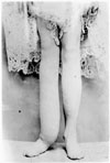
The generosity of a friend has brought a set of 12 remarkable glass plate negatives to the Cabinet. The set belonged to a Dr. Weeks from Iowa and date approximately 1890's or possibly earlier. I am going to use this opportunity to open another menu partition titled "Clinical Portraits" where all the images in the Cabinet will be indexed by subject. To begin the work, here is a portrait of elephantiasis from the Weeks set :
Finally organized a list of the photographers including a separate list of physician photographers. Something I have been meaning to do for a while.
Early views of Boston City Hospital by the photographers Allen & Rowell.
Another U. S. Sanitary Commission photo album.
With the tabloids printing news of American treatment of prisoners of war, I thought it appropriate to return to the U. S. Sanitary Commission's, Narrative of privations and sufferings... and provide an image of one of the four striking albumens that illustrate the book. A small faded snapshot displays the morbid emaciation of a soldier, and what remains of him is pale drape over a bone spindle. Against a black encroach, the light is a living presence and it has more substance than what is kited by the skeletal form. I know of no other clinical portrait that is its match for compelling our introspection. The promise of transcendence can be found in images like this, engraved by the religions of disease, even the diseases of war and hate.
Added quite a bit to the Mosso description and will leave it for now. It was fun doing the research for the book and in my readings I stumbled upon an article by Clarence J. Blake of Harvard in Boston Medical and Surgical Journal (1875) which describes a procedure for constructing a phonautograph from the human membrana tympani. The device made tracings on smoked glass with a single fiber from straw and Blake was able to demonstrate differences between the marks made by vowels and those made by consonants.
Wrote more of the Mosso description.
Still puzzling over the Mosso. Here, though is a jpeg of one of the salt prints that illustrate Kerlin's Mind Unveiled. Kerlin married Harriet Caroline Dix (b. Sep 2 1842), but I do not know if she was related to Dorothea Dix who helped out in the fund raising which this book publicized. Both women descended from Massachusetts families. I did find in Dorothea Dix's genealogy a great great gradfather, John Dix (1702 - 1787), married to a Rebecca Stone (1721 - 1786) who may also have been the great great grandfather of Kerlin's wife, Harriet. This is all just fun speculation. There is a chapter in the book signed "Humanitas" that might have been contributed by Dix, but again this is just more fun speculation.
Began the description of the Mosso with an overview.
While I slowly flog my way through the arcane Italian of the Mosso, I found a little diversion. It is a passage from an article by Dr. Laurent that was translated from the French and published in the Journal of American Insanity October 1863. Title is Physiognomy of the insane :
I saw, in September, 1860, with the greatest satisfaction, at the Stephensfeld Asylum, a room where Dr. Dagonet employs himself in taking what appear to him the most striking types of disease. M. Morel [sic] has just constructed, at St. Yon, a photographic sudio, wherein he takes the various physiognomies presented by the alienated to the eye of the physician.
In 1858, the late Dr. Ferrus, Inspector-General, caused a daguerreotype to be taken of the face of a madman who killed the much respected Dr. Geoffroy, then the chief physician. Three portraits were taken in different positions, one full face, another three-quarter, and the third in profile.
I should very much have liked to append to this essay a certain number of photographs, and thus complete, by such satisfactory proofs, the researches to which for many years I have applied myself, for pictures are more easy of comprehension than all the verbal descriptions at one's command. I must produce somewhat later this indispensable complement to the present work.
There is more to add to the Parrish description, more mysteries to solve especially the name of the photographer. I want to attribute the images to Isaac N. Kerlin, but more research is required before that can be done with any confidence.
As promised, here is a cataloging of a remarkable find by an antiquarian book dealer and scholar in Arlington. I will be devoting perhaps all of this week on the book.
From one of my sisters, here is a link to a knitted ecorche. "Its a tube isn't it?"
Decided to begin the description for the Parrish album after all.
My work cataloging the photos in the American Journal of Insanity is done for now. There is a portrait frontispiece that appeared in an 1889 issue, but the information I found comes from a bookseller's catalog and bibliographing will have to wait until I find a copy.
Before proceeding to a work by Mosso, here is an astounding portfolio of 68 albumens put together by Joseph Parrish and currently vested at the National Library of Medicine. As is the custom here, click on the date to access this work.
Put up a description for the Hun paper. I think I have one more photographically illustrated paper from the American Journal of Insanity to enter into the Cabinet bibliography and then will proceed thereafter to a work by a remarkable Italian physician.
Got an email from Patrick Pollack, an antiquarian book dealer in Devon England who found a copy of the rare Becker, Photographische Abbildungen von Durchschnitten with its 30 mounted albumen plates of the pathological eye.
I will get to the Hun description soon, but first I wanted to provide the Cabinet with this little tintype of an old woman whose profile is distorted by Bell's palsy. The opening of the mat is tiny, no more than 1 inch in width. A web search for images of Bell's palsy yielded only a handful of images, none of them from the nineteenth century, but I did find this surprisingly uplifting journal from a writer who was struck with the palsy. Also a profitable read can be found in James R. Hugunin's essay, Meditations on an Ukranian Easter Egg — I would suggest that the terrible beauty in my tintype could substitute for the demonic beauty of his Miss Winnoa cabinet card.
As is the custom here, I linked the image to an enlargement :
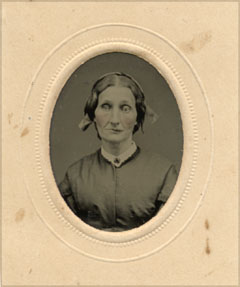
I indexed the Hun treatise and scanned in the composite photograph of ear specimens which is bound in at the end of the article. This is Hun's treatise on a rare nineteenth century asylum infection that was referred to as "insane ear."
Finished with John P. Gray articles. Will next put up another American Insanity article written by Edward Hun on haematoma auris.
Continued with the description for the second Gray article. Will try to finish it tomorrow.
Began writing the description to this second article by Gray.
This is another article by John Perdue Gray illustrated by a mounted albumen of a specimen, specifically a major portion of the skull torn off the head of a deck-hand for a coal ship. He incurred the injury from a projecting tie-rod to a bridge the boat was under passing. He was asleep at the time. The albumen is quite faded. There is a sheet of paper with an inscription attached to the specimen and after scanning at 1200 dpi and 200% I was barely able to make out the words.
Put up a description for the Gray paper.
Installed an image of Plate A for the Gray. Began reading the text.
Indexed the Gray paper.
October 10.
There are two or three more photographically illustrated papers published in the American Journal of Insanity to enter into the Cabinet bibliography. October will be devoted to accomplishing this work. Next comes a histopathology of the brain written by John P. Gray and illustrated with collotypes of photomicrographs. Gray was founder and editor of the journal and many of the anonymous editorials came from his pen.
I can not be certain how many journal articles were published with photographs. Sometimes an issue would include a card with a mounted albumen loosely inserted and I can point to one example of this, an article by Andrews entitled, Apoplexy in a Boy of Fifteen Years, which was illustrated by two seperate stereographs mounted on unbound cards. Inevitably, these artifacts jiggled free and were lost.
Completed the description for the Davies paper.
Worked on a description for the Davies paper.
Here is a curious paper by an asylum director in Britain on a case of amnesia. I doubt that there is another clinical photograph for amnesia published in the nineteenth century. I will try to read the article today and provide a description tomorrow.
Completed the description for Clarke.
Here is another treasure from the British National Library Catalog of Photographically Illustrated Books. It is a 4 page book with two mounted albumens, one of which is a collage of eleven plaster heads — phrenological models of character. Wish it were part of the cabinet! There is no information on the internet for the photographer/author E. Cruse. If you the reader can provide information, the Cabinet will be in your debt.
And another Rudanovsky.
Here is an atlas of photomicrographs prepared from nerves by Rudanovskii.
Began work on the description for the Clarke paper.
While I research the Clarke report, here is a book remarkable for its obscurity in the history of antisepsis.
It would probably take another month or two to dig up more photo-illustrated books on medical corps, but I want to move on to other subjects. October begins with this addition to the Cabinet bibliography, a composite photograph illustrating an article on "moral imbecility" appearing in the American Journal of Insanity and written by Charles Kirk Clarke, a psychiatrist who became famous for his support of the eugenics movement in North America. The photographer's name (Powell) is reproduced on two of the images but internet searches for more information are unavailing — so far. The National Library of Canada will be my next source.
Description will be provided soon.
Another U. S. Sanitary Commission publication, this one on the history of its fund raising activities that was written by Charles Janeway Stillé, a historian and patriotic pamphleteer.
Made some corrections and additions to the Belle Island report.
Finally succeeded in tracking down a description for the US Sanitary Commission report on the medical maltreatment of the Union prisoners of war at Belle Island and other Confederate prisons including Andersonville. This is an extract from the trade edition which has much more text — 283 pages of text as opposed to 52 pages in this extract. The trade edition is titled Narrative of privations and sufferings of United States officers & soldiers while prisoners of war in the hands of the Confederate authorities.
Thankyou David for the link to the British Library Catalog!
Can't find a copy of Notes and recollections of an ambulance surgeon to verify the photographic subjects, but here is the listing based on what I dug up from descriptions written by book dealers. The book is not listed by Gernsheim in his Incunabula which I find curious, although the plates are heliotypes. Certainly there must have been a presentation edition with original photographs?
Finished the description for Hospital of the Protestant Episcopal Church Began work on a book by MacCormac titled Notes and recollections of an ambulance surgeon,....
Finished the description for the Ely archive at Rochester.
Found this archive of Dr. William Smith Ely, a surgeon for the 108th Regiment of New York State Volunteers during the Civil War. Will add more biographical information soon.
Updated this important archive of Civil War surgeon, Charles, M. Clark.
Here is a listing for two albums of photographs located in the Otis Historical Archives at the National Museum of Health and Medicine. James H. Armsby graduated from the Vermont Academy of Medicine in 1833 and soon after founded Albany Medical college in 1839 when he was thirty years old.
I am reading the Hospital of the Protestant Episcopal Church and a description will take a few days. Betweentimes, here is another portfolio of clinical photographs from a Civil War hospital.
Put up jpegs of two of the albumen plates in Hospital of the Protestant Episcopal Church. They are amazingly fresh, as though taken yesterday.
Another hospital established to care for the Civil War wounded in Philadelphia. There is no author, and there is no indication of the identity of the photographer, but it is quite apparent that the six albumens are the work of John Moran. Description and jpegs will follow soon.
Here is a wonderful portfolio of images by John Moran, brother of Thomas Moran, the "dean of American painters" who is famous for his panoramic paintings of Yellow Stone National Park. The images are views of Mower Hospital which served soldiers wounded in the Civil War. The portfolio is part of the collection of The Library Company located in Philadelphia.
To honor the valiant work of governmental medical units in Louisiana, I am devoting the rest of September in a search of historical photographic works devoted to military hospitals. This history begins with the US Sanitary Commission which formed during the Civil War and involves the most important photographers of this period. Most famous is Mathew Brady, represented here by this splendid collection of 110 images documenting the Civil War. The images are all in the public domain, but not all are accessible by the Prints and Photographs Online Catalog (PPOC). I provide a link to the catalog and a call number which should be submitted as a number query, or just cut and paste the following search string : "Mathew Brady hospitals."
Finished the description for the Fluhrer.
Continued work on the description for the Fluhrer.
I was so busy researching the author of this paper, I missed the photographer's inscription on the plate. There it is on the bottom left margin, "Rockwood Phot. NY." And though disappointed that I did not discover an image by O. G. Mason, nevertheless George Gardner Rockwood is no slouch. In fact, he began his career in 1855 and was the first to make a CDV in America. He was also an early adopter of the collotype process. I am pleased to have such an eminent photographer for the Cabinet bibliography.
Began writing the description for the Fluhrer.
Fluhrer was a staff surgeon at Bellevue Hospital and this operation for gun-shot wound to the brain was performed there. For these reasons I am attributing the photographs to O[scar] G. Mason who was the house photographer at Bellevue from 1869-1909.
Subject of the four photographs illustrating the Fluhrer paper was an assistant to a butcher who shot himself in the forehead. Fluhrer is meticulous in his description of the operation and his procedures for antisepsis. Remarkably, the patient recovers with only minor complaint of memory loss.
Here is a rare offprint of a bullet injury to the brain. As yet, I am unsuccessful in discovering biographical information on the author, William F. Fluhrer. It looks as though his patient recovered from his injury, but I will read the article and provide definitive details of the case over the next few days.
Completed the description of the Epps book.
It is rare to find homeopathy texts illustrated by photographs. A first edition of this Epps with two woodburytypes of inverted nipple recently appeared on eBay so I thought it would be a good thing to share. I will put up a description soon.
As promised, here are the images.
Began the research for a monograph with two mounted silver bromides, written by Eugene Cartier for his doctorate. I have come across several of these photographically illustrated doctoral theses from France and England, but can't seem to find any from American doctoral candidates for some reason. I located two copies of the Cartier in OCLC and two in the catalogue of the Bibliotheque Nationale, three out of the four are reprints published by Baillière. I will put up the images tomorrow.
Came across this essay by Francis X. Dercum while attending the Lewis & Clark exhibition at the Mütter Museum in Philadelphia.
Here is possibly the first work by Piffard that is photo illustrated. I will add a description and images at a future date.
Finally completed a study and description of the Reverdin essay.
Here is a landmark work by Jacques-Louis Reverdin which provides some of the earliest documentation of myxoedema and other complications derived from thyroidectomy.
Found this dedication written by Edward M. Foote for his expansive tome, Minor Surgery :
This book is dedicated to the man at the point of the knife for his grit and patience, and especially for his willingness to be photographed that others may profit by his misfortune.
Of the several hundred photographs in the book, the most touching are ones illustrating a chapter on roller bandage dressing, comprised mostly of images of children.
Updated and corrected a mistake I made in describing the Bevan Lewis book on the human brain. Also scanned in the four photos.
Admittedly I am dragging out the description of the Buckminster Brown book. The book is important for any history of surgical science and, by sway of the photographic plates made by Josiah Hawes, it is also important for any history of clinical medical photography. So I am taking my time with the book. I also wanted to study the exhibition of Southworth and Hawes daguerrotypes currently on view at ICP here in New York. The Buckminster Brown book was not included in the exhibition and I have to wonder why it is so obscure. The curators, Grant Romer of the George Eastman House and Brian Wallis of ICP, put together a catalogue raisonné of Southworth and Hawes work with full size color reproductions of all the daguerrotypes in the exhibition and over 2000 additional illustrations. It is a monster of a book and I was particularly interested in the chapter titled Simultaneous Developments : Documentary Photography and Painless Surgery written by Bates and Isabel Lowry. Their essay researches the first photographic documentation of the use of ether in surgery at Massachusetts General Hospital and it was a very rewarding experience for me to have their essay on hand while viewing the five (!) iconic Ether Dome daguerrotypes in the exhibition. I question, though, how much Southworth was involved in the making of these historic images. John Collins Warren (1778-1856) is the master surgeon in the photographs and he was a devoted patron of Josiah Hawes in particular and of the new art of photography in general. It was Warren who commissioned Hawes to make the photographs of the first ether surgery and he presented Hawes with a commemorative mounting of the surgical instruments he used to perform this historic operation. Hawes made several copies of this photo, but the Getty probably has an original which is reproduced in the Lowrys' book on the Getty collection of daguerrotypes. Dr Warren is the tall figure standing on the right with his hand on the patient's left thigh. I failed to check if this image is also in the Lowrys' chapter of the catalogue raisonné.
It would have been nice to see other clinical images in the exhibition. Besides Warren, other Harvard physicians commissioned Hawes to photograph for them, especially Warren's young colleague, Dr. Henry Jacob Bigelow who with Warren's son, Jonathan Mason Warren, made all the arrangements for the first anaesthetic surgery at Mass. General.
Finished putting up the plates for the Buckminster Brown volume.
Work for the month of June begins with a book by Buckminster Brown titled Cases in Orthopedic Surgery with 8 wonderful albumens by the Boston photographer, Josiah Hawes. So far as I know, Hawes did not produce any other books with original photos tipped in.
Ending the month with this work by Anton Helwig with 16 plates of photomicrographs on toxicology. The book is quite rare and Dr. Stanley Burns graciously let me review his copy.
This will also conclude my transcriptions from Taureck for now. Her bibliography is weighted more toward works on photomicrography and there are a few volumes I did not include because they are zoological subjects, or whose illustrations are only artist copies of photographs. There are also several long descriptions of quite famous works that I did not translate, most particularly the photographic atlases issuing from the Salpetriere in Paris, and a few other books that I want to research more before putting them in the Cabinet bibliography. Certainly I will have to return to Renata Taureck's richly endowed bibliography these coming months, but in June I want to get to a few books that I treasure.
Here is an album by Ferdinand v. Heuss, famous for his "wine therapy."
Updated Kerlin.
Finished putting up images of the plates to the Rüdinger. Clicking on the thumbnails will bring up enlargements.
These are photographs of the most delicate bones of the body, tiny little convolute wonders that take in all the raucous sounds of a guttural planet and the silences beyond. Albert's photographs are magnificent, but unfortunately my camera did a poor job of rendering them for the Cabinet. I believe the Mütter Museum also has a splendid collection of temporal bones on display, including those of other species.
Worked on providing images of the plates to the Rüdinger.
Put up this atlas on the osseous anatomy of the ear by Rüdinger. A copy was auctioned off by Swann Galleries today (lot 178), the hammer price was $850.00, if I remember right. The album is comprised of 9 beautiful plates displaying the bone structure of the inner ear. 8 of the plates are large mounted ovals by Josef Albert.
Another volume on leprosy with an atlas of photographs and lithos.
Here is one of the first medical texts illustrated with photographs of leprosy. Extremely rare, only one copy found — at the Bibliotheque Nationale.
Put up this set of clinical medicine volumes by Trousseau. I did not want to forget about it. Will return, now, to the books listed in Taureck.
May 25.
This article on Dr. Stanley Burns, the distinguished historian of clinical medical photography, appeared in the Bulletin of the American College of Surgeons in January 2005 (pp. 9-15), but was posted recently on the internet. It is a PDF file but because it is only 8 pages long it should download fairly quickly.
Found this letter to the editors of the Boston Med. Surg. Journ. written by Susan Dimock while she was a medical student in Switzerland. I had to post it in the Cabinet right away.
Finished updating Maury and Duhring.
Updated Maury and Duhring.
Updated Moore on rodent cancer.
Another volume on microscopy.
Another volume on microscopy, exact same title as the Gerlach.
Updated Billroth.
Finished the Beale for now and put up this wonderful portfolio of salt prints by the Parisian photographer Auguste-Adolphe Bertsch.
Added a jpeg of the frontispiece in the Beale.
More work on the Beale. Included a link to the three page preface of the book.
Began updating Beale on the liver. Published in 1856, it is the oldest volume in the Cabinet of Art and Medicine. The plates are unremarkable — all reproductions of Beale's sketches — but the book was important for advancing the use of photography in illustrating texts on medicine. A rare and wonderful example of antique medical photography!
Finished, for now, with the Berend description.
More work on the Berend collection. Its value for the history of clinical medical photography cannot be underestimated and yet this collection is still largely unknown. Here is a link to the Wellcome Library Catalogue, key in the name "Berend, Heimann Wolff" to access thumbnails of the photographs.
Put up the citations for the Berend collection.
Began work on the Berend description. This is a collection of photographs he commissioned from the Berlin photographer L. Haase and probably intended for an atlas he was compiling. This is only a portion of the original collection, 98 photographs in all, long thought to be lost but recently rediscovered at the Wellcome Institute.
Updated the Gerlach.
Added a book by the famed Swiss orthopedist, Johannes Wildberger, which is the first German medical book to be illustrated by photographs according to Taureck.
Added the Cartier thesis listed in the catalog.
May 3.
Got a nice email from Malcolm Jay Kottler of Scientia Books who sent me a notice of a new catalog from a European dealer who is offering a few early medical volumes, notable for their photographic illustrations.
Updated Houel.
Finished the Sayre.
What a difference a shot of cortisone can make, I can work my fingers on the keyboard again! I am tabling the description for the Ross for now and returning to my work on the bibliography this week by putting up citations translated from Renata Taureck's book, Die Bedeutung der Photographie für die medizinische Abbildung im 19. Jahrundert. Her dissertation was published in 1980 and became a landmark, along with the Gernsheim, in the study of early clinical photography. I expect this work will take up most of May because of intercurrent editing and additions to the descriptions that are required. Click on the date above for a description I added to the Sayre.
April 26.
Rheumatism is slowing me down this month but the promise of warmer weather brings with it several books for the Cabinet bibliography.
Got a nice email from John Wood who is written up in a delightful article linked here »»
Worked on the description for the Ross.
Added the legends that go to the photographic plates in the Ross.
Prepared the fourth of four photo-plates that illustrate the Ross.
Prepared the third of four photo-plates that illustrate the Ross.
Prepared the second of four photo-plates that illustrate the Ross.
Put up the first photo plate that illustrates the book, A treatise on the diseases of the nervous system, by James Ross.
Commenced research on this grand opus of the Scottish pathologist, James Ross.
Added a description to the Sternberg.
Here is the plate of photo-micrographs that illustrates the Sternberg.
Began the work of adding a paper on anthrax written by Surgeon General George M. Sternberg.
Added one of Cyrus Thomson's poems to the description and will call it quits for now. I focussed on one chapter, and regret that I cannot delve deeper into the rest of the book. It really is a wonderful dark portrait of early America.
Added more to the Cyrus Thomson description and should finish it up tomorrow.
Added more to the Cyrus Thomson description.
Finished reading the Thomson book and am progressing with the description.
Put up a jpeg of the dour Cyrus Thomson cameo. It is very small, not even 4.5cm in height.
Here is a volume that is both autobiography and materia medica. Comprises wonderful vignettes of cases treated by the author, written in a folksy style.
There is enough completion to the Stewart description. Will now cast about for another book.
More work on the description for the Stewart paper, should be finished tomorrow.
Continued work on the description for the Stewart paper.
Finished reading the Stewart paper and began work on the description.
Here is a jpeg of the uranium print illustrating the Stewart paper.
Found this slim essay on obstetrics by R. Stewart. It is the first early medical text I found to be illustrated with uranium prints. Will scan in the images tomorrow.
It is very rare to find medical texts with photographic illustrations that date to the 1850's. Found this one by searching the World Catalog.
Simms description completed.
A friend of mine sent a New York Times review of Martin Parr's and Gerry Badger's The Photobook. The review is written by Philip Gefter and he covers a lot more ground than the reviewer for The Guardian who is linked below, including a mention of the photo-book, Facies Dolorosa written by Dr. Hans Killian. Gefter writes: "Another curious book, Facies Dolorosa (The Face of Pain) , was published in Germany in 1934 by Dr. Hans Killian, who photographed patients in bed and close up, labeling each image with the patient's illness. The photographs were meant for his medical research, but they show us moments between life and death with a haunting beauty that transports them into the realm of photographic art."
Added a little more to the Simms description and will try to finish it tomorrow.
Continued work on the Simms description.
Put up jpegs of the two albumens in the Simms.
February 22.
A recommended read for those interested in photographically illustrated books. Don't miss Parr's comments on Facies Dolorosa by Dr Hans Killian. It is, says Parr, 'perhaps the most melancholy photographic book of all.'
Reading A Winter in Paris and began work on the description.
Book for this week is a nice little overview of the Paris Hospitals written by an Englishman. Descriptions and images will follow in the next few days.
The final book from the Steven F. Joseph collection, on asiatic cholera by the microbiologist Van Ermengem, notable for his isolation of the bacterium Clostridium botulinum.
The third presentation from the Steven F. Joseph collection, a richly illustrated offprint on the subject of craniometry which was quite the rage in the mid 1880's!
Here is the next Belgium publication, a little gem from Henri Vercamer.
Here is the first presentation from Belgium, the Gevaert treatment for spinal deformities with plaster jackets.
February 14.
I am in contact with the distinguished photo-historian Steven F. Joseph, author of the book, "Photographers in Belgium 1839-1905" and researcher into the subject of early photographically illustrated books. Steven graciously sent to me a list of early medical volumes, illustrated by photographs and published in Belgium. I will be placing the books in the Cabinet Bibliography over the next few days and I hope some day to follow-up with jpegs of the plates when the books find their way into my library. Thank you Steven !
Here is a splendid manuscript of clinical photographs and case histories by a Civil War Surgeon. It is a long forgotten gem owned by the University of Chicago Library. I hope it will be published some day!
Completed the description for the Stevens book and will proceed to the next book.
Plate VI is done.
Plate V is done.
Plate IV is done.
Plate III is done.
Began work on the description to "Funtional Nervous Diseases." I will be adding Steven's text that corresponds to each of the six double before-and-after portraits. Plates I and II are done.
Scanned and uploaded all the image files to "Functional Nervous Diseases".
Here it is on page 21 that Stevens gives his extraordinary premise for the book: "Difficulties attending the functions of accommodating and of adjusting the eyes in the act of vision, or irritations arising from the nerves involved in these processes, are among the most prolific sources of nervous disturbances, and more frequently than other conditions constitute a neuropathic tendency."
Note to self: cure the disease by curing how the disease is perceived!
Began reading the Stevens textbook. The book does not appear to be that rare - located about 24 copies in American libraries.
January 25.
Finished work on the Keith paper. Next book to be added to the Cabinet bibliography will be "Functional Nervous Diseases" by George T. Stevens.
More work on the description of Doctor William Keith's paper, including a link to a drawing of the projectile which he removed surgically.
Began the description of the Keith extract.
Added a jpeg copy of the albumens to the Keith extract. Before and after images of the repair made by Keith. Am unable to find much of anything from the life of William Keith other than an account book located in the archives of Aberdeen University, Scotland.
After a considerable amount of bother, all the files of the Cabinet – some 1400 in number – are now transposed onto a new computer.
Work for the year of 2005 will begin with a description of this rare work by the surgeon William Keith. I believe it holds the earliest published photographs for plastic surgery, specifically the surgical treatment of a traumatic skull injury.
Back to work on the bibliography. Returning to the Humphry volume, I corresponded with the photo historian who attributed the plates to Valentine Blanchard and he kindly sent to me jpegs of the femurs in his copy of the book. Inscribed under the plates is the following text : "Valentine Blanchard & Lunn, Cambridge."
January 1.
This journal entry is posted from a borrowed computer, a Sony laptop with keys the size of the heads of 16 penny common nails. I am looking forward to a good year in the hunt for knowledge and a new computer.
