Nach dem franzˆsischen mit Autorisation des Verfassers deutsch bearbeitet und durch zahlreiche Zus‰tze erweitert von Berthold Benecke.
Braunschweig : Friedrich Vieweg und Sohn, 1868.
Description : [i]-xiv, 265 p., [1 l.] errata, [2 l.] pl. & [2 l.] tbls. ; ill., 2 phot., 88 figs. ; 23 cm.
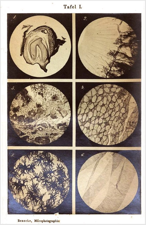
Photographs : 2 composites each with 6 photomicrographs and each facing a table of descriptive letterpress.
Photographer : Berthold Adolf Benecke.
Subject : Photomicrography.
Notes :
Cited :
Taureck, Renata, Die Bedeutung der Photographie für die medizinische
Abbildung im 19. Jahrhundert (1980) ; p. 204 :
The textbook has two photographic plates as well as 88 wood cuts illustrating the text. It is an edition of a textbook published by A. Moitessier in 1866 and enhanced by Benecke's own experiences. B. Benecke (1843-1886) was very much involved with the technical advancement of photomicrographic instruments and recognized their value for microscopic anatomy. Here he depicts composited tissue preparations, "that often photographically depict isolated elemental organisms, but that are less suitable for showing the achievements of photomicrography."
The pictures are well executed, e.g. tissue cuts from the skin of the human head (25 : 1), transverse strip of m.gastrocnemius muscle bundle of frog (50 : 1). Diatome photographs in incremental enlargements (800, 1000 and 2000) that were achieved by progressive enlargements of the original negatives and that are quite rough, but not so unclear as comparable examples from Reichardt and Sürenberg, likewise from 1868.
Ich beschloss daher anf‰nglich mit Aufgabe meiner eigenen Arbeit dies Werk [of Moitessier] einfach zu ¸bersetzen und meine Erfahrungen in Form von Zus‰tzen und Anmerkungen hinzuzuf¸gen, kam aber bald zu der Ueberzeugung, dass es zweckm‰ssiger sein w¸rde, meine Resultate nebst denen deutscher und englisch - amerikanischer Forscher mit dem franzˆsischen Werke organisch zu verbinden. Deutsche und ausl‰ndische Journale wurden mir zu diesem Zwecke in Menge zur Disposition gestellt, auch erhielt ich noch im vorigen Jahre die vierte Auflage von Beale's Handbuch der Mikroskopie zeitig genug, um part IV, On taking photographs of microscopic objects (p. 229ó279) f¸r meinen Zweck benutzen zu kˆnnen.
In dem vorliegenden Werke schliesst Moitessier's Arbeit mit dem sechsten Capitel der zweiten Abtheilung ab, doch hoffe ich, dass auch aus den folgenden Abschnitten, bei deren Bearbeitung namentlich die grossen Werke von Hardwich, van Monckhoven, Barreswil und Davanne u. A. benutzt wurden, der angehende Mikrophotograph zweckm‰ssige und fˆrderliche Belehrung schˆpfen werde.
Die beiliegenden Probebilder habe ich nach ‰lteren Pr‰paraten mit den in Fig. 35 und 22 abgebildeten Apparaten aufgenommen und theilweise mittelst des Apparates Fig. 74 noch vergrˆssert. Die zu einer Tafel zusammengehˆrigen, auf gleichstarkem Glase hergestellten Negative wurden dann passend zugeschnitten und durch Colophoniumkitt und schmale schwarze Papierstreifen auf einer Spiegelplatte befestigt und zusammen copirt. Die Copieen sind in dem Atelier des Herrn Prothmann unter meiner Controle angefertigt und nach einigen Versuchen sehr gleichm‰ssig ausgefallen. Jede Retouche ist sowohl auf den Negativen, als selbstverst‰ndlich auch in den Papierbildern unterblieben. Bei der Auswahl der abzubildenden Objecte habe ich vorzugsweise zusammenh‰ngende Fl‰chenpr‰parate ber¸cksichtigt, da isolirte Elementarorganismen schon ˆfter photographisch abgebildet wurden und weniger geeignet sind, zu zeigen, was man in der Mikrophotographie zu leisten vermag.
Und so ¸bergebe ich denn dieses Handbuch den deutschen Forschern mit dem Wunsche, dass es beitragen mˆge zur Fˆrderung der Mikrophotographie und der mikroskopischen Erkenntniss.—Pages viii-ix.
At first I decided to simply translate this [Moitessier's] work with examples of my own labors and follow it up with my experiences in the form of comments and additions, but quickly came to the conviction that it would be much more appropriate to connect organically my results—as well as those of german, english and american researchers—with the french works. Piles of German and foreign journals were put at my disposal, also, in the previous year I acquired the fourth edition of Beale's manual on microscopy in sufficient time to be able to use part iv, On taking photographs of microscopic objects (p. 229ó279), for my purposes.
In what follows, Moitessier's work closes with the sixth chapter in part two, however I hope that the ensuing excerpts, especially those adaptations from the great works of Hardwich, van Monckhoven, Barreswil and Davanne and others that will be used, will make for appropriate and helpful instruction for the prospective photomicrographer.
The accompanying test shots I took from old preparations with the apparatuses pictured in figures 35 and 22 and some of them enlarged even further by means of the apparatus in fig. 74. For a composited plate, a selection of equal thickness glass negatives were produced and then cut to size and fastened with colophon glue and small black paper strips to a mirror and photographed together. The copies were prepared in the studio of Mr. Prothmann under my control and after a few experiments, quite consistent. As is understandable, all retouching is in the negatives and does not extend beyond to the paper images. In the choosing of photographable objects, I have considered preferable relevant surface preparations, since elementary organisms have been depicted photographically quite often and are less likely to show what one can carry out with photomicrography.
And so I now deliver this handbook over to german researchers with the wish, that it may contribute to the advancement of photomicrography and the knowledge of microscopics.