The results of information received respecting nearly nine hundred persons who had attained the age of eighty years, including seventy-four centenarians.
Cambridge : Macmillan and Bowes, 1889.
Description : front., xii, 218 pp., 2 pl., 1 db. pl. ; ill., 4 pl.s ; 19.5 cm.
Photographs : 3 woodburytypes [1 port. front.].
Photographers : Valentine Blanchard and Colin Lunn.
Subject : geriatrics, statistics.
Cited :
Goldschmidt and Naef, "Truthful Lens", figure 24.
Notes :
Reprinted, augmented, from the "British medical journal" and the "Collective investigation record". Includes Annual oration delivered to the Medical Society of London, 1885.
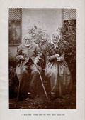
FRONTISPIECE: Photograph of Benjamin Atkins and his wife, each of whom was 101 (see page 208).
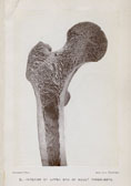
Of the following PHOTOGRAPHS the first shows the section of the Upper End of the Thigh-bone of an adult man, the form of the part, the angle of the neck with the shaft, and the structure of the interior. The wall of the bone, enclosing the medullary cavity, is seen to be thick below, to diminish in thickness as it ascends into the neck in consequence of plates separating from it which form the cancelli of the neck, head and great trochanter. A vertical series of these cancelli ascend to the uppermost part of the head ; they are the chief recipients of weight from the pelvis, and serve to transmit the weight from that part to the inner side of the wall of the shaft. Others form an arch subtending the upper wall of the neck and transmitting weight from it to both sides of the wall of the shaft.
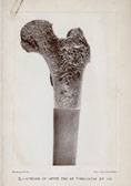
The second shows a similar section from a woman reported
to have died at the age of 103. There is no reduction in the size
or alteration in the form of the bone. Indeed, for a woman, it
is large, and the neck forms an open angle with its shaft. But it
contrasts with No. 1 remarkably in the thinness of the wall of the
bone, which has resulted from absorption at the interior, and in the
diminution of the cancelli with the consequent enlargement of the
spaces between them. The bony plates which form the arch subtending the
upper wall of the neck can scarcely be recognised, and
the vertical series ascending to the summit of the head are much
reduced in number and size.
The contrast thus presented is sufficient to account for the fact
that fracture in this region is common in old persons — especially in
women, the angle of the neck with the shaft being at all periods of
life more open in women than in men — though it is comparatively
rare in adults and young persons.