The College of Physicians of Philadelphia.
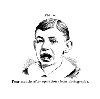
The set of four photographs of a grimacing elfin waif appearing on pages 144 and 145 of the Mütter book is captioned, "Unidentified disease or disorder of the jaw." The child was a patient of Dr. J. Ewing Mears, a distinguished professor of anatomy and clinical surgery at the Pennsylvania College of Dental Surgery. The images were reproduced as woodcuts to illustrate his paper titled, Closure of the jaws and its treatment published in the College of Physicians Transactions (Fig.s 4, 5, 10 & 11) 32 The child's inability to close his jaw was the result of cicatricial and tissue damage from a cancrum oris.
The photograph falling on page 203 of the Mütter shows a specimen of fetal encephalocele, delivered intact by Rohrer and discussed by Charles Delucena Meigs in his work, Obstetrics, the science and the art. 33 A woodcut illustration appears twice in that work, on pages 225 and 430. Following is the excerpt:
Here is a repetition of the figure of Dr. Rohrer's case, given at 224. Let the Student observe that such a vast fluctuating tumor on the vertex of a child could never be the really presenting part; that it must necessarily deviate, and go up in the iliac fossa, allowing the true head to present, and making that, of course, a face presentation. I say of course, for the head would be of course in extension. Well–the labor going on–the head is born in face presentation–giving the face the appearance of suggillation–of which I have made a good representation in the figure; but the head being born, the tumor, larger than the head, remains above the strait in the same way as the second head of the double-headed fœtus of Dr. Pfeiffer's did. Here, then, is a case in which evolution is indispensable, and Dr. Rohrer informed me that this was what he brought about–after doing which, he was enabled, with very violent force of traction, to pull away the caput succedaneum–as you see it in the figure.
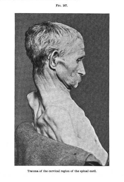
Wood's photograph on page 141 appears as Figure 167 illustrating a chapter on diseases of the spine that Lloyd contributed to Dercum's A text-book on nervous diseases. 34 The photo also illustrated a paper written by Lloyd and published in volume XIX of the Journal of Nervous and Mental Disease in 1894. 35 The disease represented is muscular atrophy caused by inflammation to the subject's dura mater—in this case the symptoms of hypertrophic spinal pachymeningitis, first described by Charcot in 1871.
The case of acromegaly shown on page 80 of Mütter looks to my eye like a younger version of the subject shown on page 895 of the Dercum textbook (Fig. 294), illustrating Joseph Collins's chapter on trophoneuroses.
The photograph featuring a wax model of the famous Madame Dimanche with her horn appears on page 67 of the Mütter. A similar photograph was used by Buffalo, N.Y., surgeon Dr. Roswell Park to illustrate his paper titled "Diseases and injuries of the head," published as a chapter in the Dennis surgical compendium. 36 The half-tone appears on page 509 of this book as Fig. 386 and is captioned "Cornu cutaneum (Wood Museum)." Following is Park's description:
Another form of scalp-tumor of not very rare occurrence is the so- called " cornu cutanenm " ( Fig. 386). This is a veritable cutaneous horn formed by circumscribed hypertrophy of the epidermis, and consisting of an outgrowth of horny consistency and appearance and of variable size and shape. These horns are usuallv tapering, maybe either straight or curved, and occasionally are completely bent upon themselves Sometimes they assume flattened forms; at other times they are but slightly elevated. In color they are of a dirty, grayish-yellow appearance, although when old and exposed to accumulations of dirt from outside sources they may be found almost black. Their surface is rough, irregular, more or less fissured, and the ends may have various shapes. As a rule, the horn scarcely ever is more than a half an inch in diameter at the base, which latter rests directly upon the skin. The adjacent tissues are usually normal, there being an abrupt transition from healthy skin to the base of the outgrowth. Sometimes, however, large papillae are found in the neighborhood. They are for the most part solitary ; still, multiple horns have been recorded. In a case recorded by Batge1 the whole lower part of the body was studded with these growths. While they may occur elsewhere, they are by all means most common upon the scalp and face. They usually occur late in life, but have been observed in the young. They are hard, dry, more or less brittle ; are rarely painful, except when disturbed. They are slow in growth, and frequently loosen and drop off, but are almost always reproduced. They seldom disappear spontaneously. They bear close resemblance to warts, and, in fact, are essentially hypertrophic warts. As in the case of nearly all cutaneous outgrowths, they may be followed by epitheliomatous degeneration. The only satisfactory treatment for these growths is complete excision of the entire mass, with extirpation of the area from which they grow, including the periosteum.
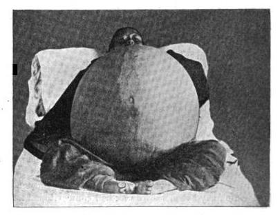
This photograph, as well as the two stunning cabinet cards appearing on pages 186 and 187 of the Mütter, were taken in 1894 to illustrate Dr. Elizabeth Reifsnyder's paper titled, Some remarks on abdominal surgery in China; with report of removal of large cyst of left ovary (Figs. 1, 2 & 3 vide: »»). 37
Yü Yung Né, aged 25, married, and a farmer by occupation, was admitted October 22d, 1894. When I first saw her she was standing outside the hospital, resting her tumor on a wheelbarrow. She lived some forty-five miles distant; had come with her father in-law to see if she could be cured. Former patients had told her of our hospital, and so the painful journey was taken. I told her father-in-law I could not promise that she would live, to which he replied : " No matter; go ahead and cure."
Coolies carried her in; she was weighed—two hundred and forty-six pounds—and then put to bed. History as follows: As a child always healthy ; menstruated at 15 ; periods painless and always regular until two months ago, when they ceased ; was married at 19; never had any children—in fact, is little more than a child herself; apart from her tumor, about the size of the average American girl of 13.
Reifsnyder's photographs of Yü Yung Né were also reproduced as half-tones in Gould and Pyle, Anomalies and Curiosities in Medicine (pages 783 & 784 of the 1898 London edition).
32.) Mears, J. Ewing, (1887), Closure of the jaws and its treatment.
Philadelphia: "Transactions of the College of Physicians of Philadelphia" ; pages 69-81.
33.) Meigs, Charles Delucena, (1856), Obstetrics, the science and the art.
Philadelphia: Blanchard & Lea pages; pages 225-226, 429-430.
34.) Dercum, Francis X. (1895), A text-book on nervous diseases by American authors. Philadelphia: Lea Brothers & Co.;
pages 538-539.
35.) Lloyd, James Hendrie (1895), Diseases of the spinal cord. A text-book on nervous diseases by
American authors (Chapter XVIII); Philadelphia: Lea Brothers & Co.; pages 538-539.
36.) Park, Roswell, (1895), Diseases and injuries of the head. Philadelphia: Lea Brothers & Co.;
from: "System of surgery"; vol. iii., pages 497-786.
37.) Reifsnyder, Elizabeth, (1895), Some remarks on abdominal surgery in China; with report of removal of large
cyst of left ovary. New York: "The American journal of obstetrics and diseases of women and children" ; vol. xxxi., pages 512-520.