The College of Physicians of Philadelphia.
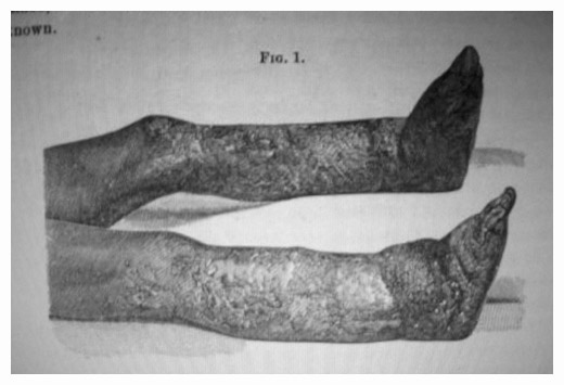
Perhaps third in historical importance is the photograph of Dr. James Buckner Luckie's case of synchronous triple amputation falling on page 63. At a time when the mortality rate of a single amputation at the thigh was 61 percent, Luckie's patient survived the amputation of both legs at the thigh and almost the entirety of his right arm. This was the second successful triple amputation in the United States, and the Mütter description includes the astounding fact that Luckie accomplished the first successful synchronous triple amputation as well! The museum owns both historic images and published the photograph of the precedent case on page 47 of the first Mütter volume. Luckie wrote up a report on both cases in a paper titled, Two cases of successful simultaneous triple amputation for railway injuries,10 published in the Fort Wayne journal of the medical sciences which was promptly picked up by several other leading journals. The Fort Wayne journal was devoted exclusively to railroad injuries and was edited by Dr. Christian Berry Stemen, president of the of the National Railway Surgeons' Association, and it was before that "great army" that Dr. Luckie from Birmingham, Alabama read his reports at a meeting in Chicago, June 28, 1888.11 Steman also included Dr. Luckie's reports in a chapter on amputations in his book of railway surgery.(ibid.) Here in its entirety is the second report:
Case II.—On the morning of December 11, 1865, I was called to the Pioneer Company's Works, three miles from the city, to see J. McKnight, white, æt. 32 years, who had been crushed under the wheels of a car loaded with building material. Upon questioning the messenger I found that the injuries were very similar to those in the case reported above, and that probably a similar operation would have to be performed. I at once sent for Dr. Copeland, who so ably assisted me in the former case, and requested him to accompany me. Upon arriving at the house where the wounded man lay, I found both legs horribly crushed up into and a little above the joints, and the right arm crushed into a mass from the tips of the fingers to above the elbow.
Some one had given him a dose of morphia and a stimulant, and he was resting passably well; pulse a little feeble, but regular. Assisted by Dr. Cunningham, of Pratt Mine, and such help as we found present, we at once made arrangements to operate. Dr. Cunningham administered the ether. As soon as the man was thoroughly etherized we applied our Esmarch bandages and operated as in Case I, Dr. Copeland taking the left and myself the right side. Dr. Copeland amputated the left thigh at its middle third, whilst I amputated the right at the juncture of the middle and lower third. So simultaneously did we amputate that after I had sawed through the right femur I did not lay down the saw, but reached over and sawed the left for Dr. Copeland. After tying the vessels I then amputated the right arm at its middle third. We dressed the stumps as quickly as possible and placed the man in bed.
He came from under the influence of the ether badly, and for a while we had grave fears as to the result. He, however, rallied and steadily improved day by day. He convalesced without any trouble whatever, except from the stump of the left thigh; upon removing a button of dead bone all trouble was remedied. The operation the same as in Case I, the modified circular. The man is now living and is hale and hearty.
On page 133 there is a picture of elephantiasis in a seated male subject which I believe was made by the photographer Christiano Junior for his atlas Casos Notables de Elefantiasis, durante su permanencia en el Brasil sometime in the late 1860's, if memory serves me. 12
On the opposite page is another elephantiasis image from the same period which is attributed to Dr. A. Newman. The patient depicted is John P. T., age 34, who was treated surgically by Dr. Thomas G. Morton (1835-1903) at the Philadelphia Hospital, for the first time on December 12, 1873 with ligation to his femoral artery and a second time five years later with an excision of a portion of his sciatic nerve. The second operation was unprecedented, an experiment based on the observation that damage to the nerve trunk led to atrophy in an affected limb. Following is Morton's reasoning behind the procedure, extracted from his book, Surgery in the Pennsylvania Hospital/ 13:
His condition was so distressing that he was anxious for an amputation of the limb. The case now seemed out of the reach of any surgical procedure. Thinking over the fact, prominently brought to mind by several cases of wounds of nerves where atrophy had followed nerve-section, it occurred to me that a division or section of the trunk of the sciatic nerve might be followed by such change in nutrition and subsequent diminution in size as to give the patient in this case some relief; his condition being as to warrant such an experiment, even if the limb were to a great extent palsied, it would be better than the risk of such a serious operation, without considering the deformity from amputation, which, as the disease existed at least as far as the middle of the thigh, would require to be made very high up on the thigh. I also recalled the fact that a section of the sciatic would not necessarily involve a permanent loss of usefulness of the limb, for the nerves would, doubtless, in time reunite, and, even if the progress of the disease were not permanently arrested, much would be gained.
The second operation had dramatic results and the limb lost much of its mass, but the patient died five months later from complications of pneumonia and an abscess in the thigh according to Morton's report. More likely, he died from sepsis. It is a thrilling narrative that would have enhanced the Mütter volume if it had been included. Had Morton's neurectomy cured this dreadful disease, then this photograph would be better known. Instead it is almost a cognate of Tuskegee and an uncomfortable reminder of experimental medicine's past indiscretions. Pictured above is the wood engraving that Morton used to illustrate the case.
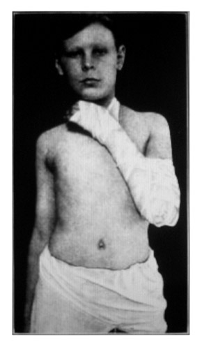
On page 45 is a set of sequential photographs on the technique of roller bandaging for maintaining hyperflexion in the treatment of fracture of elbow. It should be noted that the term hyperflexion of the elbow is identical to the "Jones position" discussed by Astley Paston Cooper Ashhurst in his book An Anatomical and surgical study of fractures of the lower end of the humerus. 14 The photographs were made for this work, appearing on pages 89-90, and in his prefatory remarks Ashhurst writes, "The photographs, made by the author, have been skillfully retouched by Mr. Charles F. Bauer," so now we know the name of the author/photographer. It should also be noted that Dr. Ashhurst's treatise won the Samuel D. Gross Prize of the Philadelphia Academy of Surgery in 1910 and that the photos were used again by Ochsner for his 1922 monograph.
I counted 6 photographs of Civil War subjects, each of them excellently captioned. All images are from the eight volume set, Photographs of Surgical Cases and Specimens: taken at the Army Medical Museum (1866-1881) by George A. Otis (1830-1881).15 Page 19 shows two photographs from a set of three, numbered 298, [299 not included] and 300 that come from Volume VI. Page 23 photo is number 262 from Volume VI. Page 27 photo is number 289 from Volume VI. Page 28 photo is number 282 from Volume VI. Page 29 photo is number 93 from Volume II. A seventh Civil War subject appears on page 179 of the Mütter, the preparation of a transverse colon which came from a soldier who died of dysentery. The case was reported on page 512 of Medical and Surgical History of the War of the Rebellion/ Medical Volume Part Second and the plate itself faces page 514 of that work.
Page 31 holds a spectacular image of a recovered trauma patient with cicatrices from a stab wound extending the length of his thorax. The patient was treated by Dr. Augustus V. L. Brokaw who reported the case in the St. Louis Courier of Medicine under the title, A unique case of stab wound of thorax and abdomen: recovery.16 The remarkable recovery became a testimonial for a product called Antikamnia which Brokaw used instead of opium to treat pain.
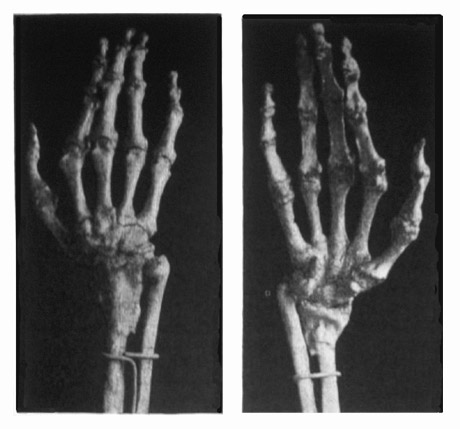
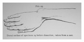
What follows is a little more explanation for the images on pages 38 and 39. The two photographs of the plaster cast were among a set of at least 6 photographs that Dr. Roberts received from Dr. Alfred Scott of Dublin. Two of the photographs showed the skeletal remains represented by the cast, and were reproduced in his paper titled, A clinical, pathological, and experimental study of fracture of the lower end of the radius, with displacement of the carpal fragment toward the flexor or anterior surface of the wrist17 published in the Transactions of the American Surgical Association in 1896 and as a separate offprint in 1897. Roberts gives the following explanation:
Bennett refers in his paper to a similar specimen " from the museum of this college, to which it was presented by Mr. Swan in 1885." He gives no description of the specimen, and evidently means by his remark that it is in the Museum of the Royal College of Surgeons in Ireland. I have obtained, through the courtesy of Mr. J. Alfred Scott, of Dublin, photographs of this specimen, showing the palmar and dorsal surfaces of the bones. He has also sent me photographs of the plaster cast taken of the forearm and hand before the soft parts were removed; and plaster casts of the forearm and of the lower ends of the radius and ulna. There is no clinical history of the injury. It is probably a disecting-room specimen.
The term Smith's Fracture used for a forward displacement of the radius is named after the Irish surgeon Robert William Smith, (1807-1873) and was thought to be quite rare until Roberts was able to document more cases than anticipated. J. Alfred Scott also published papers on Smith's fracture. Scott was a man of dextrous intellect and also published on the mathematics of photography, so there can be little doubt that he was the maker of these images. I have not been able to find the source of the skiagraph shown on page 39, but my guess is that it was used by Dr. Roberts' for his 1901 publication on Smith's fracture. I also conjecture that the provenance of the cast and skeleton might be found with Sir Astley Cooper (1768-1841) who I believe wrote the first published description, but this is probably over-reaching. Above are the other Scott photographs and below is a drawing of the cast, all taken from the Roberts book:
10.) Luckie, James Buckner, (1888), Two cases of successful simultaneous triple amputation for railway injuries.
Fort Wayne: Journal of the medical sciences; pages 164-166.
11.) Stemen, Christian Berry, (1890), Railway surgery; a practical work on the special department of railway surgery;
for railway surgeons and practitioners in the general practice of surgery.
St. Louis: J. H. Chambers & Co.; pages 186-188.
12.) unverified: Christiano de Freitas Henriquez Junior, José, (ca. 1866), Casos Notables de Elefantiasis, durante su permanencia
en el BrasilRio de Janeiro : sn, sp.
13.) Morton, Thomas George (1880), Surgery in the Pennsylvania Hospital; being an epitome of the practice of the Hospital since 1756; including
collations from the surgical notes, and an account of the more interesting cases from 1873 to 1878; with some statistical
tables. Philadelphia: J. B. Lippincott & Co.; page 115.
14.) Ashhurst, Astley Paston Cooper, (1910) An anatomical and surgical study of fractures of the lower end of the humerus.
Philadelphia: Lea & Febiger; pages 89-90.
15.) Otis, George Alexander, (1866-71), Photographs of surgical cases and specimens; taken at the Army Medical Museum,
Washington, D. C. Washington: Surgeon General's Office.
16.) Brokaw, Augustus V. L., (1890), A unique case of stab wound of thorax and abdomen; recovery. St. Louis: The
St. Louis courier of medicine; vol. iii, 257-263.
17.) Roberts, John B., (1896), Clinical, pathological, and experimental study of fracture of the lower end of the radius,
with displacement of the carpal fragment toward the flexor or anterior surface of the wrist. Philadelphia: Lippincott;
"Transactions of the American Surgical Association"; vol. xiv, 611-682, 1 pl.