The College of Physicians of Philadelphia.
The caption for the image of Osler on page 220 requires further annotation for the slower medical students who have picked up this book. Osler was the greatest clinician of his day and this image has the radiance of a touchstone, smudged and damaged from the generations of medical students who have pulled it from the archives to touch the legend. Osler was an outside talent brought in to occupy the chair of clinical medicine at UPenn, vacated by Dr. William Pepper (secundus) in 1884 and the picture shows him in his natural element as a consummate anatomist in the Blockley dead house — known alternately as the "green room" or "green house." He performed over 160 autopsies at Blockley and each session packed the room with medical students — even the skylight above was used for viewing. The one woman in the group who is pictured on the right is Amelia Gilman who later served as physician to the Blockley Insane Asylum.
Regarding the photograph of the green room on page 218, I would like to learn more about the painting hanging on the left wall.
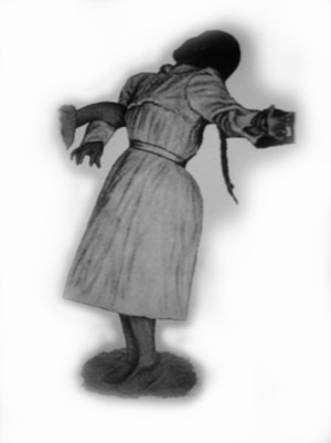
The photographs on pages 194 and 195 are captioned as "unidentified mental illness," but without a doubt they represent a case of astasia-abasia, or Blocq's disease, discussed by Lloyd in Dercum's A Text-book on Nervous Diseases. 18 His essay is titled "Hysteria" and a photograph very similar to the two in the Mütter can be seen on page 124 of the Dercum. Following is Lloyd's description of the case:
M. R. aged, twenty-eight years, German, was a patient in my wards at the Methodist Episcopal Hospital. She had a history of convulsive attacks and of several spells of ataxia. Five weeks before admission she had been badly frightened. Then followed a series of hysterical prodromes, such as fulness of the head, auræ in the stomach, pulsations in the chest and throat, and headache. Then followed an abortive seizure. Then the astasia-abasia developed suddenly. This was the most marked symptom for some weeks. It is illustrated in the photograph (Fig. 39). The patient required support either side. The movements of the legs were wildly inco-ordinate, tending now to fly outward, now to be drawn up, and again to be arrested by a tonic contracture, which drew the patient up on her toes, with her body backward as in opisthotonos. But little forward progression was made unless the patient was urged or partially carried along. When sitting or lying she had full control of her legs, and full power in them. The knee-jerks were exaggerated. The patient had other stigmata, the most marked of which was an almost total anæsthesia, as shown in Fig. 28.
The name of the photographer who took the brain photo that falls on page 191 is Charles Trought, of the Army Medical Museum in Washington. Dr. Daniel Lamb with Dr. Woodward (1833-1884) both led the post-mortem and Lamb also participated with the team appointed to perform the gross examination of the Guiteau brain. First, however, the brain was sent from Washington Jail — where it was extracted — over to Trought's studio for the photo session. Following is what Lamb reported about the circumstances of the photographing19:
The external appearances of the body should first be noted ; the cavities, except the spinal, should be opened and their contents examined; that after the removal of the brain, and its examination without incision, it should be transferred, properly guarded and protected, to the Army Medical Museum, where, through the courtesy of the Curator, Surgeon D. L. Huntington, U. S. Army, it would be photographed, and then a cast be taken ; that its internal structure should then be observed, and portions set apart for microscopical examination ; and that the entire operation should be completed, as far as possible, the same day, to enable physicians resident elsewhere, present by invitation, to return promptly to their homes; and that the notes should be taken in duplicate.
By reason of delays, for which neither I nor my assistants were responsible, the examination was not begun until 2:30 P. M. (one hour and a half after death), in consequence of which the photographing was less successful, and a cast was impracticable.
Pages 157-159 show three photographs by Ambrose H. Lincoln of Boston. These are original plates from Howard Franklin Damon's (1833-1884) photographic atlas of skin diseases published in 1869-1870 with the title, Photographs of Skin Diseases, Taken from Life, Under the Superintendence of Howard F. Damon.20 The work has the distinction of being the first photographic dermatology text published in the United States. Following are the captions that go with the photos in the order of their appearance in Mütter:
PLATE II. Herpes circinata is occasionally accompanied by herpes iris; and it is more frequently associated with herpes tonsurans. They are diseases of childhood, and occur on the exposed or unprotected surface of the body. Thus, the face neck, and extremities are the seats of election for herpes circinatus and iris ; while the hairy portions of the scalp are attacked by herpes tonsurans. The photograph represents herpes circinatus of the forehead, and herpes iris on the cheek of a little girl. These diseases have with good reason been attributed to alterations in the nutrition of the skin and its appendages, produced by the presence of a parasitic plant, the tricophyton tonsurans.
PLATE XII. The form of zoster, commonly known as "shingles," occupies a lateral half of the thorax, and terminates at the median line in front and back. A similar half-zone occasionally appears on the abdominal region. Zoster is preceded and accompanied by neuralgia of the part. This is often the most troublesome feature of the disease. The eruption consists of rosy patches, often the size of the hand, oval, circular, irregular, and confluent, studded with numerous straw colored or pinkish vesicles, varying in size from a mustard-seed to a large pea. These become mature during the first week, dessicate during the second, or, being ruptured, form brownish crusts, which fall off at the end of the second, or during the third week. The pain usually subsides after the formation of the vesicles. Sometimes ulceration takes place. The distribution of the vesicular clusters is upon those regions of the body supplied by the terminal branches and filaments of the principal peripheral or cutaneous nerves. Zoster may be divided into the following varieties: Zoster facialis, cervicalis, brachialis, thorachialis, abdominalis, lumbalis, and sacralis. Zoster frontalis, nasalis, labialis, auricularis, and præputialis, are subordinate varieties of this cutaneous neurosis.
Plate VII. Dermatologists have employed various names in order to distinguish the different conditions and appearances assumed by chronic eczema. Thus, it is known, in its dry stage, as eczem siccum and squamosum; and as eczema fissum, rimosum, and fendillé, when there are fissures or chaps in the skin. This form of eczema is peculiar to washerwomen and those whose hands are frequently immersed in soap and water. The epiderma is removed so rapidly and so often, in some instances, that the derma becomes irritated, infiltrated, and loses its natural elasticity: chaps or fissures are thus formed. These are quite painful, and occasionally bleed.
Although the disease topic represented by the photograph on page 193 is unknown, it is consistent with Dercum's photographic experiments with induced epileptoid seizure. I would wager there is an unpublished manuscript somewhere, incomplete because it is missing this photograph. This image, especially, of all the mystery photographs in the Mütter book, is the one that I find most intriguing and want to learn more about.
Page 81 is a case reported by Morton with the title, Two cases of congenital hypertrophy of the fingers published in the Philadelphia Medical News.21 Morton amputated the affected fingers:
DR. Thomas S. K. Morton made some remarks upon the subject of congenital hypertrophies of the extremities and reported two marked cases of such hypertrophies of the hand. The first case occurred in a child aged 18 months. The index and middle fingers of the right hand were enormously hypertrophied in all their elements, while the remainder of the hand was normal. Almost no function existed in the affected fingers. Amputation was resorted to, successfully, at the metacarpo-phalangeal articulations. Microscopic examination showed that all elements entering into the diseased fingers were hypertrophied by a fibro-fatty degeneration. The joints were but poorly evolved.

The beautiful photograph of vascular protrusion in the left orbit which is shown on page 32 is the case of Dr. George C. Harlan who published his report in the American Journal of the Medical Sciences in 1870 under the title, Traumatic aneurism of orbit, treated by compression.22 His report follows a paper on orbital aneurysmal disease by Thomas G. Morton who also discussed Harlan's patient and provided a woodcut reproduction of the photograph.23 What follows are excerpts from Harlan's report:
S. G., śt. 25, a brakesman on Pennsylvania Railroad, was admitted to the Wills Ophthalmic Hospital on the 14th of August, 1869. On the 20th of June, 1868, while standing on the platform of a car in motion, he received a violent blow upon the head from another car moving in the opposite direction on a side track, which fractured the lower maxilla on both sides and inflicted several scalp wounds. He stated that he was partially insensible for three weeks, and that when he returned to consciousness his right eye was very prominent and swollen, and that he heard "a roaring sound like falling water in his eye" with every stroke of the pulse, which he had never been free from since.
At the time of his admission there was great exophthalmos of both eyes—greater of the left which seemed almost dislocated, the equator of the ball being in front of the edge of the orbit. The left ball could be pressed back to its proper position and easily held there without pain to the patient, but immediately bulged forward again when the pressure was removed. The right eye resisted pressure, its position could not be altered, and strong pressure gave pain. He had excellent binocular vision.
On consultation of the surgeons of the hospital, it was decided to give a thorough trial to treatment by compression of the carotid, as the pulse was full and strong, and excited by prolonged examinations and the prospect of surgical treatment – beating at the rate of nearly 100 per minute – tinct. veratrum viride was given several hours before compression was commenced, and repeated at intervals. The resident surgeon, Dr. James C. Wilson, assisted by Mr. Landis, an advanced student of medicine, and the patient himself, who was an intelligent man of unusual endurance and indomitable perseverance, kept up continuous and complete compression from 3 P.M. until 11 P.M. – a period of eight hours – the pulse ranging in the mean time, under the influence of the veratria, from 44 to 56.
This treatment was continued for five weeks, during the last two of which no progress was made. Bather for the sake of confirming the assurance that all had been done that could possibly be accomplished by pressure, than with any decided hope of further improvement, continuous pressure was tried, being kept up by relays of students for twenty-four hours, but with scarcely any effect. The patient then left the hospital, expressing himself satisfied that the degree of improvement attained fully compensated him for all he had endured. A few weeks ago he wrote to me to say that he had resumed his occupation as brakesman, and that the former condition of his eyes had returned, that he was no better than when he entered the hospital. He will probably return to undergo the operation of ligation of the carotid.
18.) Lloyd, James Hendrie, (1895), Hysteria. Philadelphia: Lea Brothers & Co.; "A Text-book on Nervous Diseases
By American Authors/"; pages 87-134.
19.) Lamb, Daniel Smith, (1882), Report of the post-mortem examination of the body of Chas. J. Guiteau, (who died
by hanging, June 30, 1882, at the United States jail, Washington, D. C., in execution of judicial sentence).
Chicago: Chicago Medical Press Association; "The Chicago medical journal and examiner"; vol. xlv., pages 73-78.
20.) Damon, Howard Franklin, (1870), Photographs of Skin Diseases, Taken from Life, Under the Superintendence of Howard
F. Damon. Boston: James Campbell.
21.) Morton, Thomas S. K., (1894), Two cases of congenital hypertrophy of the fingers. Philadelphia: Penfield
Publishing Co.; "The Medical and Surgical Reporter"; page 291.
22.) Harlan, George C., (1870), Traumatic aneurism of orbit, treated by compression. Philadelphia: Henry C. Lea;
"American journal of the medical sciences"; vol. lx., pages 46-48.
23.) Morton, Thomas George, (1870) Orbital Aneurismal Disease, and Protrusion of the Eyeball from Venous Obstruction;
with Remarks and Cases. Philadelphia: Henry C. Lea; "American journal of the medical sciences"; vol. lx., pages 38-46.