Journal : Revue photographique des hôpitaux de Paris ; vol. 1.
Paris : Adrien Delahaye, 1869.
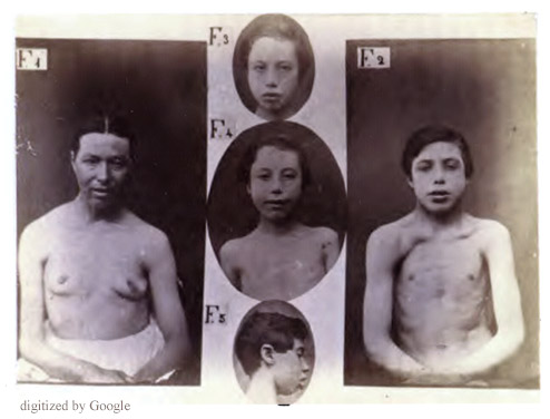
Description : [1 l.] pl., 41-45 p. ; ill.: 1 photo. ; 24.5 cm.
Photograph : mounted albumen, composite of 5 images.
Photographer : Duchenne.
Subject : Skeletal muscles — Duchenne muscular dystrophy ; Dx.
Notes :
Les cinq figures photographiques qui forment la planche IX, représentent le thorax et la face de sujets atteints d'atrophie musculaire progressive de l'enfance. — L'atrophie musculaire progressive, lorsqu'elle fut décrite pour la premiére fois, était considérée comme une maladie exclusive à l'àge adulte.—Page 41.
Une autre affection de l'enfance, beaucoup plus rare que la paralysie atrophique graisseuse, la paralysie pseudo-hypertrophique, récemment décrite par M. Duchenne de Boulogne, a été confondue par un pathologiste anglais, M. Edw. Mergon [sic], avec l'atrophie musculaire de l'enfance.—Page 45.
This paper supplemented a series of articles Duchenne published in the Archives Générales de Médécine [1] on the juvenile form of hereditary progressive muscular dystrophy that he named paralysie musculaire pseudohypertrophique and paralysie myosclerosique (Duchenne pseudohypertrophic muscular dystrophy). With these terms Duchenne classified pseudohypertrophic muscular dystrophy as arising strictly within the skeletal muscle anatomy and thereby distinguished the disorder from childhood paralyses caused by brain stem or spinal degenerations and lesions such as SMA (spinal muscular atrophy) and polio. Duchenne was the first to publish on the differential diagnosis of the childhood progressive myopathies and he was the first neurologist to use the camera as a diagnostic tool for this purpose.
Duchenne photographed the four subjects depicted here who include three members of the Hottmann family, the mother (Fig. 1) and her two sons aged 9 and 14 (Fig. 2, Figs. 3-4) all of whom he diagnosed with atrophie musculaire progressive [de l'enfance], his term for paralysis caused by hereditary motor neuron degeneration (Duchenne-Aran muscular atrophy). Adults are usually affected, but for the Hottmann family, facial paresis and eversion of the lower lip occurred in childhood and were the first signs of disease process. This fact led Duchenne to mistakenly believe they belonged to a rare subgroup, de l'enfance, that he thought he had discovered and first identified in the 1861, 2nd edition of De l'Electrisation localisée— case observations CI and CIV (pp. 453 and 478). [2] He photographed case observation CIV and published two of his images of the patient in an atlas complementaire to the textbook (vide: Fig. 1 »» & Fig. 2 »») along with a third profile portrait of a boy with the same diagnosis (Fig 16 »»), [3] a younger version of the boy portrayed here in the composite as Fig. 5.
His error is then aggravated with the false claim that it was this cohort of spinal disease that Dr. Edward Meryon (1807–1880) was describing in a paper that is now recognized by most scholars as the first classical account of pseudohypertrophic muscular dystrophy. [4] Confusion over the pathogenesis of myofacial paresis appears in numerous reports of the time and, unfortunately, Duchenne contributed to the confusion with this paper and his refutation of the work of Meryon.
Charcot, who championed Duchenne's work and helped him prepare the histological evidence for the third edition of De l'Electrisation localisée, incorporated Duchenne's paper in a lecture titled, Révision nosographique des amyotrophies, in which he wryly noted that "Duchenne's disease" must be rare for it was hardly mentioned in the standard literature! [5] The following is an excerpt from the Charcot lecture where he points out Duchenne's error:
Passons maintenant à une autre forme que Duchenne (de Boulogne) a décrite comme représentant simplement une variété de l'atrophie musculaire progressive et à laquelle il a donné le nom de forme infantile de l'atrophie musculaire progressive. Elle semble être rare, car elle n'est guère mentionnée dans les traités classiques. Duchenne, dans son Traité de l'Electrisation localisée, dit en avoir rencontré une vingtaine de cas, et, dans la Revue photographique des hôpitaux, on trouve des photographies, faites par Duchenne lui-même, et représentant la face de plusieurs malades atteints de cette affection. Ici, le début de la maladie se fuit, suivant la description de Duchenne, par la face et en particulier par l'orbiculaire des lèvres. Les lèvres se renversent en dehors de manière à simuler l'aspect habituel de ces organes chez les strumeux. Les membres ne se prennent que consécutivement, les bras d'abord, puis le tronc. Enfin, fait important à signaler, cette forme infantile est héréditaire et l'on peut retrouver dans une même famille des parents atrophiques engendrant des frères et des sœurs atteints à leur tour d'amyotrophie débutant à la face. D'après tout ce qui précède, on serait tout naturellement porté à conclure que l'amyotrophie relève, dans ces cas, d'une lésion spinale, comme dans les cas du type Duchenne-Aran, dont ils ne représenteraient, suivant Duchenne lui-même, qu'une simple variété. Ces prévisions seraient cependant mal fondées. MM. Landouzy et Déjerine ont en effet présenté, l'an dernier à l'Académie des Sciences, l'histoire de cas typiques de l'atrophie musculaire progressive in fantile de Duchenne, et dans un de ces cas, l'aulopsie a démontré qu'il n'existait aucune lésion, soit de la moelle épinière, soit des nerfs périphériques. Il s'agit donc encore dans ces cas de myopathies primitives.
Duchenne believed that facial paralysis in children could only occur in non-pseudohypertrophic forms of hereditary muscular paralysis, but this theory was finally disproven—as noted by Charcot—by the work of Landouzy and Dejerine in 1885 (v. intra: »»). Facies myopathica, childhood onset, asymmetrical atrophy of muscles and reversal of the anterior axillary fold—especially pronounced in the 14 year-old Hottman brother—all fit the clinical picture of fascioscapulohumeral syndrome of muscular dystrophy established by these two authors. Returning to the boy portrayed as Fig. 5, he was unrelated to the Hottmann family, but represented, according to Duchenne, another example of his mystery cohort. His name was Henri Juliard and when Duchenne first photographed him for his Atlas complementaire he was 13 years old. It is the first image published of Landouzy-Dejerine syndrome.
1 Duchenne, G. B. (1868), Recherches sur la paralysie musculaire
pseudohypertrophique ou paralysie myo-sclérosique, Paris: P. Asselin;
"Archives Générales de Médécine," 6. s., xi. (5, 179, 305, 421, 552).
2 Duchenne, Guillaume-Benjamin (1861), De l'Electrisation localisée et de son application
à la physiologie, à la pathologie et à la thérapeutique, Paris: J. B. Baillière et fils, 1861 (2nd edition).
3 Duchenne, Guillaume-Benjamin (1862), Album de photographies
pathologiques complementaire du livre intitulé De l'electrisation localisée, Paris:
J. B. Baillière et fils; Plates 1, 2 & 16 v. intra: »».
4 Meryon, Edward (1852), On granular and fatty degeneration of the voluntary muscles.
London: Medico-Chirurgical transactions, Volume 35 ; pp. 73-84 & pl., vide:
»».
5 Charcot, Jean Martin (1872-1887), Leçons sur les Maladies du Système nerveux
faites à la Salpêtrière. Recueillies et publiées par Bourneville, Paris: A. Delahaye & E. Lecrosnier; page 192.