Image source: Google books (August 27, 2020: »»)
Leipzig : Verlag von F. C. W. Vogel, 1877.
Journal : Archiv für Ohrenheilkunde ; vol. 13, issue 1.
Description : 26–68 p., [1 l.] pl. ; illus: 12 phot. figs. ; 24 cm.
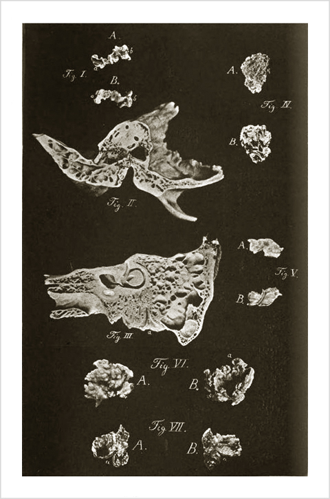
Photograph : collotype (Lichtdruck). Composite photograph of specimens, Figs. I, IV-VII with front and back views.
Subject : Mastoid — Chronic mastoiditis ; perforations & sequestra.
Notes :
Fig. 1. Sequester aus dem rechten Antrum mastoideum mit der knöchernen Scheidewand zwischen Antrum und Sulcus sigmoid. Von der Innenseite A gesehen zeigt er bei a die glatte cylindrische Wand des Sulcus sigmoid. mit seinen Gefässlöchelchen, im übrigen eine rauhe unregelmässige Fläche, welche durch Loslösung von der Pyramide entstanden ist (die obere Kante des Bildes stellte in situ die untere dar). Von der Aussenseite B gesehen erscheint eine glatte, mit mehrfachen grösseren Löchern versehene, leicht concave Fläche, die innere und theilweise untere Wand des Antrum mast. mit den Einmündungen der Warzenzellen, bei a die Scheidewand zwischen Antrum und Sulcus, bei b das rauhe verdickte Ende, welches theilweise dem innersten Theile des Gehörganges, theilweise der hinteren Paukenhöhlenwand angehört haben wird.
Fig. 2. Horizontaldurchschnitt des Schläfenbeines der in der Höhe der Spina supra meatum durch Gehörgang, Paukenhöhle und Antrum mast. gelegt ist; in das letztere ist der Sequester Fig. 1 B in seiner ursprünglichen Verbindung mit schwarzem Contur eingezeichnet.
Fig. 3. Verticalschnitt des linken Schläfenbeines durch die Crista superior des Felsenbeines gelegt, zeigt das Verhältniss der spongiösen Substanz in Pyramide und Warzentheil zu den pneumatischen Räumen. Bei a ist das For. stylomast. in den Schnitt gefallen
Fig. 4. Sequester der vordern äusseren Partie des Warzentheiles angehörig von einem vierjährigen Knaben. A Aussenfläche, B Innenfläche mit den Scheidewänden der Warzenzellen.
Fig. 5. Nekrose der vorderen Hälfte des rechten Os tympanicum von einem siebenjährigen Mädchen. A Aussenfläche, B Gehörgangsfläche mit dem Sulcus pro tymp., nach links vom Sulcus dem Gehörgang, nach rechts der knöchernen Tuba angehörig. (Die untere Kante von B war in situ die obere.)
Fig. 6 und 7. Concentrische Epidermoidalmassen von Fall II. Fig. 6: A äussere bucklige, den erweiterten Warzenzellen entsprechende Fläche, am Präparat von weisser Farbe mit starkem Perlmutterglanz, B concave Innenfläche dieser Masse mit deutlicher concentrischer Schichtung, bei a rosettenförmig angeordnet. Fig. 7 der usurirten hinteren Wand des Gehörganges von Innen anliegende Lamelle; A auf ihrer concaven dem Antrum, B auf ihrer convexen dem Gehörgang zugewendeten Seite. Bei a ist in A und B noch der vermeintlich in den Gehörgang gemachte Schnitt sichtbar. —Page 67-68.

Bezold's habilitation thesis, the second in his corpus on mastoid disease. His first study was a statistical analysis of the odd anatomy of the mastoid and the implications for surgical perforation procedures (cf. Bezold: »»). Here he investigates perforations caused by sequestra in both carious and necrotic mastoid disease, pursuant to untreated and long standing inflammatory processes, typically arising within the middle ear (otitis media). To prepare for his thesis, Bezold compiled 111 cases of diseased temporal bones of which 34 involved the mastoid and congruent parts, and 42 involved the mastoid alone. He breaks the n42 subset down to 15 caries, 16 necrotic caries, and 11 necrotic cases. The n34 subset is broken down to 20 caries, 10 necrotic caries, and 4 necrotic cases. He notes that the n34 subset is often symptomatic of a destructive constitutional pathology such as tuberculosis, scrofula, or syphilis, but still produced sequestra 14 times greater in the mastoid. Sequesters were even more prolific in the n42 subset, numbering 27. These numbers raised the question that Bezold sought to answer: what is it in the nature of the mastoid that conduces resorption and osteonecrosis?
Anamnesis is carefully documented for four of his patients: Ernst W.–age 6, Postal official F.–age 34, Anna V.–age 8, and Mrs. H.–age 34. A fifth case of a 7-year-old girl is mentioned, but Bezold refers the reader to her report in an earlier issue of the journal (1870: »»). The clinical picture is completed for three of the patients by front and back photographs of their sequestra. Figure 5 is a necrotic fragment of os tympanicum removed from the external acoustic meatus of the 7-year-old girl. Figures 6 and 7 are two examples of the bowl shaped layered epidermoid masses evacuated from Postal official F.'s mastoid process. Figure 1 is the sequestrum of Mrs. H., measuring 15 mm. long by 5 mm. wide, removed after advancing forward into the auditory meatus. Bezold was surprised to see that 1 cm. of her sequester was smooth on both sides, and deduced that it broke off the septum between the sigmoid sinus and the antrum. Proof is found in Figure 2, representing a horizontal slice of temporal bone with the puzzle piece outlined in black, perfectly matching Mrs. H's bone fragment.
Bezold proposed aggressive cleansing and debridement of the ear canal at the first signs of mastoid disease and treated his patients with Listerian spray, later introducing boracic acid in the treatment protocol (1880: »»). Elements of the terms, "Bezold's sign" and "Bezold's abcess," are inchoate on pages 51-52, but entered the classical medical literature with his lauded third treatise of the mastoid corpus, "Ein neuer Weg für Ausbreitung eitriger Entzündung aus den Räumen des Mittelohrs auf die Nachbarschaft und die in diesem Falle einzuschlagende Therapie" (1881: »»). In that work he used colored gelatin on cadavers to trace the paths of mastoid empyema from an abcess in the medial plate emptying into the digastric groove and beyond. Bezold continued to publish shorter clinical reports on mastoid disease but his final synoptic work in the corpus is a pedagogical chapter in the Schwartze "Handbuch der Ohrenheilkunde," with an illustrated anatomy followed by updated descriptions of the primary infections (1893, vol. ii, p. 299-351: »»)
