The College of Physicians of Philadelphia.
...nor is there any better way to advance the proper practice of medicine than to give our minds to the discovery of the usual law of nature, by careful investigation of cases of rarer forms of disease.
Nor could there be a better way to open a book of historic medical photographs than with the quoting of William Harvey (1578-1657) from a letter he wrote to Dutch physician John Vlackveld on April 24, 1657.1 Vlackveld had sent Harvey information on the postmortem of a plasterer, whose diseased bladder had an unusual viscus no larger than "a walnut with the husk" and walls "the thickness of the little finger."
As a rule, only cases of rarer forms of disease came to be photographed by the first physician photographers of the nineteenth century. Even so, there were very many medical mysteries to record in the 19th century and it is surprising that the Mütter has less than a dozen daguerreotypes of clinical subjects in its collection. Just as few in number reside at the Warren Museum in Boston. This scarcity can be blamed on an acquired notational bias among physicians, namely, that photographic make-ready is disposable once it is published. In the possession of these physicians from the age of heroic medicine, even expensive cased images lost their preservation value once they were reproduced as woodcuts or engravings in the literature. It is not surprising, then, that the photographs in museum repositories like the Mütter are often disembodied from their provenance with only a few scribbled notes penned on the back by a reporting physician's illegible hand writing (another acquired notational bias!).
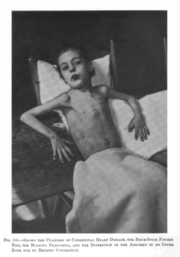
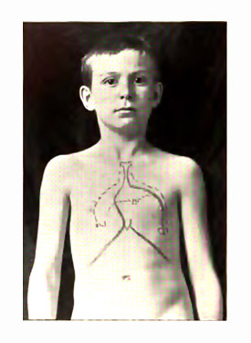
A project of restoring lost provenance to photographic artifacts like the ones presented by the latest Mütter Museum release is a formidable challenge. Case in point: the compellingly poignant image of a young girl on page 123 whose chest is a map of cardiac and hepatic regions of dullness. The only other clues that remain to this historical image are her name, "Jennie Savage," and the caption, "Children's Hospital, taken about 12/15/????" (possibly 1894). Eyes looking at the picture are immediately drawn to the pronounced bulge along the left edge of her sternum that could be caused by hypertrophy in the right side of the heart, or possibly by fluid in the pericardium, or maybe even a mediastinal condition. If Jennie has a stenosis, is it acquired or congenital? A congenital mitral deficiency is usually immediately detectable at infancy with unmistakeable signs such as the distinctive cyanosis that evokes the term "blue baby." Jennie however, is pale, not cyanotic, and shows no signs of respiratory distress. And if this really were a case of a long standing mitral stenosis, why didn't the doctor pose her for the appropriate clinical picture of this condition, taking care to position her fingers in a way that would show signs of clubbing, and baring more of her midriff to show any abdominal distension? The 1903 Babcock 2 has a classical illustration of congenital mitral stenosis:
The diagram of dullness painted on Jennie's chest, enormously proportioned even for a child, just adds more confusion because it is closer to the dimensions of pericardial effusion than it is of an enlarged heart as demonstrated by this illustration, from a later edition Rotch 3 :
Further confirmation of pericardial effusion would be a lower location of apex beat, even as far as the 6th
intercostal but still within the area of dullness. In the 1890's when Jennie's photograph was taken and before xray
technology could be used as a diagnostic tool in cases of cardiovascular disease, auscultation alone divined the hidden
contours of the heart. In the picture of Jennie could that be an "x" that is barely perceptible, marking the location
of apex beat inside the lower angle of dullness? Is her bulge more typical of effusion than it is of an enlarged heart?
Heart problems were not uncommon in the pediatric wards of hospitals at the time — a frequent complication of many childhood diseases including tuberculosis, rheumatic fever, pneumonia and even less virulent diseases such as measles. I assumed that there had to be something rare and exceptional about Jennie's case that warranted the making of a photograph, and spent a frustating day looking through medical library catalogs for an acquired pediatric stenosis or a case of paracentesis, or any unusual case of pediatric heart disease published in Philadelphia. A report of Jennie by John Bingham Roberts (1852-1924) would have been a great find, because he was an important early advocate of the often fatal technique of paracentesis for aspirating the pericardium in last-ditch cases. Plus, he was a Philadelphia man, but this search turned out to be a dead end. So too did my searches for strange cases of pericarditis and mitral stenosis coming out of Philadelphia within the time period of 1899-1901 when a report would have been published, assuming the Mütter reference is correct. Could the Mütter date be wrong? On the photo it is so faint it looks as though it were erased. Or perhaps it is another "Children's Hospital" in another city where she was treated, maybe Chicago or London?
I did eventually find Jennie and her doctor, not where I expected to find them, but still within the Children's Hospital of Philadelphia and close to the time indicated by the photograph. Her story is included in the closing section of the text that follows which is intended as a bibliographical guide to the Mutter Museum Historic Medical Photographs. I hope you will read this with an appreciation for the problems of sourcing the material. It is a nearly impossible task considering the lack of research funds and the stretched resources of this great grey lady cadaver of Philadelphia, but that it was a labor of love for Laura Lindgren, the editor and co-author, is clearly evident. The book is a bright prospect for biblioforensicists of medical photography who obsess over this material because we can now expect that it will prompt other great repositories of medical photographs to come forward with their collections — Musée de l’hôpital Saint-Louis, Warren Anatomical, do you hear?
1.) Willis, Robert, (1847) The works of William Harvey, M. D. London: Syndenham; pp. 616-617.
2.) Babcock, Robert Hall, (1903) Diseases of the Heart and Arterial System. New York, London: Appleton & Co.; page 691.
3.) Rotch, Thomas Morgan, (1903) Pediatrics: The Hygienic and Medical Treatment of Children. Philadelphia:
J.B. Lippincott Company; page 760.