Prepared, in accordance with the acts of Congress, under the direction of Surgeon General, Joseph K. Barnes, United States Army.
Washington: Government Printing Office, 1879.
Description: xii., 869 pp., 41 plates, 44 figures, 30cm.
Second printing: also 1879.
Photographs: 41 heliotypes or woodburytypes.
Subject: Civil War, 1861-1865, war medicine.
Photographers: E. J. Ward.
Cited:
Garrison & Morton, #5185: Garrison considers this the greatest
single monograph on dysentery. Woodward saw the Lösch
amoeba, but without recognizing its significance; he was part
author of the Medical and surgical history of the War of the
Rebellion.
Notes:
• The medical history vols. were prepared by J. J. Woodward,
Charles Smart and George A. Otis.
• The surgical history vols. were
prepared by George A. Otis and D. L. Huntington.
• Both Otis and Woodward died before the publication dates
of the surgical volume Part III and the medical volume Part III.
For an excellent synopsis of Woodward's editorial involvement in this encyclopedic medical publication read "An enduring monument": Philadelphia's contributions to The Medical & Surgical History of the War of the Rebellion (1870-1888), by Michael Rhode Society for the History of Authorship, Reading and Publishing »».
The following description of the plates appears in the second issue:
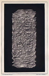
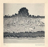
"NOTE — Of the foregoing illustrations the colored diagrams, Plates I and II, were executed by Mr. E. Sinclair, of Philadelphia; the chromos, Plates III, VI, VIII, XIV, XIX, XX, XXVII, XXX, XXXI, and XXXIII were executed by F. Moras, of Philadelphia, under the supervision of Mr. Hermann Faber, by whom the original water-color drawings were made. Plates IV, V, VII, XV, XVI, XVII, XVIII, XXI, XXII, XXIII, XXVIII, XXIX, XXXII, XXXIV, XXXV, XL, and XLI are reproduced from photographs of the specimens selected, made at the Museum by Mr. E. J. Ward. With the exception of Plate VII they were reproduced for the first five thousand copies of this work after the Woodbury-type process by the American Photo-relief Company, under the supervision of Mr. John Carbutt, of Philadelphia. This process, commended by Mr. ALEXANDER AGASSIZ — (No. 2, "Applications of Photography to illustrations of Natural History," Bulletin of the Zoological Museum of Cambridge, November 30, 1971,) and subsequently employed by that naturalist for a portion of the plates of his splendid "Revision of the Echini," Cambridge, 1872-73 — was also selected by my colleague, Assistant Surgeon G. A. Otis, U. S. A., for a number of the photographic plates in the Second Surgical Volume of this work. Meanwhile, however, the Heliotype process had been made available in this country by the enterprise of James R. Osgood & Co., of Boston, under the supervision of Mr. Ernest Edwards, and as this method was found to give results which in delicacy and beauty closely approximate the Woodbury-types, while the plates, being executed in printer's ink on ordinary plate-paper, must be as permanent as the letter-press itself, it was selected for the second five thousand copies authorized by the act approved June 23, 1875. Plate VII was reproduced by the Heliotype process for both editions. Plates IX, XXV, XXVI, and XXXVI are copied from photo-micrographs, magnified 12 diameters, by Assistant Surgeon E. Curtis, U. S. A. They were etched on steel by Mr. H. Faber. Plates X, XI, XII, XIII, XXIV, XXXVII, XXXVIII, and XXXIX are fac-similes of photo-micrographs made by myself, with powers varying from 63 to 280 diameters. They were reproduced for the first five thousand copies by the Woodbury-type, and for the second by the Heliotype process, as in the case of the other photographic plates. The original negatives, and the preparations copied, are preserved in the Army Medical Museum."
J. J. WOODWARD, Surgeon, U. S. A.
However it should be noted that the comment on Plate VII may be a typesetter's error because the image appears as a woodburytype in my second issue edition, not as a heliotype.
Although Woodward parses an overwhelming amount of statistics in this magnum opus, the number of deaths caused by the alvine fluxes in the Federal Armies was still an approximation at 57,265, so enormous was the devastation worked by the pathogen Entamoeba Histolytica. For example, another 15,000 troops were mustered out of the army for "debility" which no doubt included a sizable percentage of fatal cases of diarrhoea. Thousands more continued to die after the end of the war and up to 14 years after discharge. However, these numbers are a much smaller percentage of the the number of soldiers who fell sick but were able to recover during the term of their service. Woodward writes:
[These disorders]...made their appearance at the very beginning of the war, not infrequently prevailing in new regiments before their organization was complete, and although comparatively mild at first were not long in acquiring a formidable character. Soon no army could move without leaving behind it a host of the victims. They crowded the ambulance trains, the railroad cars, the steamboats. In the general hospitals they were often more numerous than the sick from all other diseases, and rivaled the wounded in multitude. They abounded in the convalescent camps, and formed a large proportion of those discharged from the service for disability.
Woodward gathered his information from forms circulated among doctors serving both the Union and Confederate armies. Andersonville Prison, for example, reported a disportionately larger percentage of chronic cases ending in death, 5605 out of 7352. Overall, colored troops fared better with the ratio of chronic to acute forms of dysentery and diarrhoea, but suffered a worse prognosis for death. Mortality for the colored troops, averaged 1 out of every 83 cases of acute diarrhoea, and 1 out of every 4 cases of chronic diarrhoea, whereas deaths for white troops averaged 1 out of every 395 cases of acute diarrhoea and 1 out of every 6 cases of chronic diarrhoea.
The plates are beautiful images of mostly diseased colons, some ilia cut lengthwise and stretched and tied to glass rod frames. Woodward made the splendid photomicrographs and presented them for display at the U.S. Centennial International Exhibition in 1876 prior to publication.