1 PERFORATING GUNSHOT FRACTURES OF THE SKULL. -- A few instances were reported in which men survived after
perforations of the craniums by musket balls. The abstract of the following ease furnishes an instructive example,
the more interesting because the patient has been kept under observation for nearly nine years from the date of the
reception of the injury:
2 CASE. -- Private Patrick Hughes, Co. K, 4th New York Volunteers, aged 23 years, was wounded at the battle of Antietam,
September 17th, 1862, in Sumner’s attack on the right, near Dunker’s Chapel. Several special reports, at variance in regard
to some particulars of the case, have been received. In the note book of Surgeon J.H. Brinton, U. S. V., it is stated that
“two missiles had struck the back portion of his head, the one near vertex causing injury of greatest extent,” and that
“he did not entirely lose his consciousness at time of injury.” But the field report, and the majority of the subsequent
hospital reports, state very positively that the injury was a perforation of the skull by a single conoidal musket ball
entering near the inner posterior angle of the right parietal, and emerging at a higher point of the left parietal,
making, after traversing a portion of the brain, a large exit wound. Little is known of the early history of the case,
except that the patient dragged himself from the field, and, after a preliminary dressing from Surgeon G. W. Lovejoy
of his regiment, was conveyed to a field hospital in a barn near Keedysville, where he remained until the 20th,
when he was sent to Hagerstown. The regimental surgeon reports that the shock and depression from the injury was great,
but that the patient was conscious and answered questions rationally. There is no report from the Hagerstown hospital,
except that the patient was sent to Washington on the 24th. On the 25th, he was received at the Mount Pleasant Hospital.
The following is an extract from the report of Acting Assistant Surgeon Thomas Carroll, who had immediate charge of the
case, after the patient’s admission to Mount Pleasant Hospital: “The ball entered half an inch posterior to the junction
of the coronal and sagittal sutures on the median line, passed backward under the scalp and escaped one inch above the
occipital protuberance, inflicting a wound four inches in length, producing a compound comminuted fracture of the skull
of the same extent. At the time of his admission to the hospital, eight days after the reception of the wound, so much
tumefaction of the parts existed that it could not he ascertained whether or not the brain or its membranes were injured.
The general condition of the patient was good; suppuration had commenced; no febrile action existed, the pulse was regular,
sleep not materially disturbed, mind clear and manifesting no signs of compression of the brain, or inflammation of its
membranes. Little, if any, change was perceptible for several days, when the swelling of the scalp and tissues subsided,
leaving a prominence nearly, if not altogether, one inch in height, and two and a half or three inches in length of brain
substance, in which the pulsation of the arteries could be distinctly observed. From the closest examination that could now
be made, it was supposed that the membranes of the brain were not lacerated, though this supposition was by no means
certain, as there was reason to believe the brain itself lead been penetrated. The chief, and in fact, the only unpleasant
symptom complained of by the patient, was an occasional paroxysm of giddiness that occurred at intervals of from ten to
fifteen days; the paroxysms continued to return, though less frequently, while he remained in the hospital. Early in
December, he was able to sit up and walk about the ward, and was never afterwards much confined to bed. This protuberance
now began to subside, and was soon reduced nearly to a level with the skull; numerous pieces of bone were removed as they
became detached from the tissues, leaving a complete channel in the cranium from the point where the ball entered to where
it emerged. At the time this man was discharged from the service, January 26th, 1863, the wound was nearly healed. There
was but little discharge of pus, and with the exception of an occasional recurrence of the paroxysms of giddiness, he
suffered but slight inconvenience. The treatment consisted simply in cutting the hair short, and applying cold water dressing locally,
paying strict attention to the diet, and administering a cathartic as often as symptoms seemed to
indicate its use.” Assistant Surgeon C. A. McCall, U. S. A., in charge of Mount Pleasant Hospital, furnished a report of
the case, from which the following extracts are made. “The ball passed from about one inch and to the right of occipital
protuberance into the cavity of cranium and emerged at a point on the left of the median line, about one and three-fourths
inches from the point of entry; probably a conoidal ball. From the posterior margin of the interior wound to anterior margin
of superior was about four inches. At the time of his admission to hospital, eight days after the injury, so much tumefaction
and hardened clot existed, that it was deemed inadvisable to make a very strict examination with a view to determine the
extent of lesion. The hair was closely shaved, and cold water dressings applied and patient placed in a sitting posture.
The general condition of patient was promising, although all the brain appeared clouded, the memory impaired, speech
desultory or given in fragments, yet being readily aroused and, generally at such times, answering promptly and clearly.
In a short time healthy suppuration was established, febrile action decreased, the pulse became regular, sleep became
natural, the mind began to show clearness, and he seemed to lose all symptoms of compression or of inflammation of the
brain or its membranes. The swelling and induration of the parts gradually disappeared, leaving their place a fungus
cerebri of considerable size, perhaps an inch in height by two amid a half or three inches its length, in which the
pulsations could be distinctly seen and felt. The only unpleasant symptom now existing was the occurrence of a paroxysm
of giddiness at intervals of ten or twelve days, which symptom continued, although at much greater intervals, during
the remainder of his stay at the hospital. The size and extent of the fungus deterred me from using the knife, and I
had resort to compression as recommended by the surgeons of France, but with such serious results that I was startled
for the safety of my patient, and therefore threw aside all dressing save the simple cerate out soft lint, and proper
guard for the delicate tissues. Early in December, he was able to sit up and walk about the wards, and was never
afterwards confined to the bed. From this time the fungus commenced to decrease in size, and continued to do so until
shortly before his discharge from the service, January 26th, 1863, when it had shrunk below the level of the skull,
and both openings had been well covered by cicatricial tissue. During the progress of the ease numerous spiculae of
bone were removed as they became detached. The constitutional treatment consisted in paying strict attention to the
diet, and exhibiting saline cathartics and turpentine injections as the symptoms seemed to call for. When the soldier
left my hands he complained of no inconvenience whatever with the exception of the occasional attack of giddiness, and
the fact that he could not see a small object when placed in a position directly on a level and on a middle line between
the eyes. Both which defects, I consider, will diminish with time until lost. Shortly before the patient’s discharge
from Mount Pleasant, an excellent picture of the aspect of the injury at that time was made in water color, under the
direction of Surgeon J. H. Brinton, U. S. V., who was then in charge of the Division of Surgical Records of the Surgeon
General’s Office, and had secured the services of an artist, Mr. Stanch, whose admirable drawing and coloring have
furnished some of the best illustrations of this book. The figure on the right of the chrome-lithograph opposite is a
good copy of Mr. Stanch’s water-color drawing. I take the following memorandum of the case from one of the memorandum
books turned over to me by Dr. Brinton: “Gunshot wound of head: The picture of this case was taken at the Surgeon
General’s Office, whither the man came as an orderly, or on an errand. He was perfectly well. A small carnified cerebral
hernia existed at the exit wound, which was healing rapidly. The ball went into cavity of the skull and emerged. No
symptoms existed at all, when the patient’s portrait was taken. The wound was then some three or four months old. The
history of this case, which I took at the time, has been unfortunately mislaid.” After this we have the history of the
case taken up by the pension examining surgeons. The patient went to Newcastle, Delaware, and was pensioned at four
dollars a month, until June, 1869, when his pension was increased to eight dollars a month. Dr. D. W. Maull reports,
at this date, that there was a loss of substance of the skull two inches wide and three inches long, leaving a large
depression, covered only by time integuments of the cranium. “Through this can be seen at all times the pulsations of
the brain. About the cavity the bone is ridged by the union of the fractured margins. There is almost constant dull
pain, some loss of hearing, and the sight of the right eye is impaired.” Dr. Maull regards the disability as total.
Yet the man worked afterwards in an iron foundry as paddler. On December 20th, 1870, this patient was examined by
Doctors William Thomson and W. W. Keen of Philadelphia, who have published an interesting account of his condition
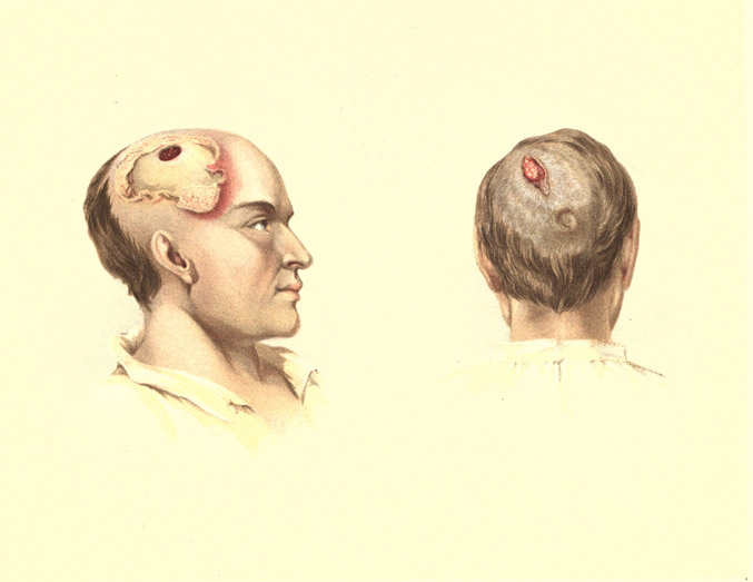
at that time, with a photograph, of which a reduced copy is given in the annexed wood-cut, (FIG. 106.)1
I make the
following quotations from this paper: “His memory is quite good, but by no means so good as before the injury. He
is rather easily bothered and confused, and more irritable than formerly. The sight of his right eye, he thinks, is
poor. Whisky affects him as usual. Sexual power undiminished. He has no paralysis. The wound of entrance is marked
by a slight depression in the bone, the wound of exit by a hollow two and a half by two inches, and one inch deep. No
bone has closed this opening, but the scalp and hair dip down into the hollow. The arterial pulsations are barely
perceptible. When recumbent the hollow is gradually obliterated and replaced, in about one minute, by a rounded
protuberance. To prevent pain during this change, he supports the parts with his hand. When he coughs, even with
moderate force, the depressed scalp instantly hedges up in a cone, which nearly reaches the general level of the
skull and obliterates the depression, and then as suddenly subsides.” Drs. Keen and Thomson observe that “the complete
recovery from paralysis, (as evinced by his subsequent severe labor,) and the almost entire restoration of his mental
faculties, are remarkable, especially in view of the probable deep lesion of the brain, both by the primary injury and
the subsequent fungus cerebri.” They comment also upon “the rapid changes in the state of the cranial contents, due to
any change of position, to coughing, etc., as evinced by the effacement of the depression at the wound of exit.” The
authors also regard the case as of value in throwing light upon the probable anatomy of the optic commisure. Their
discussion on this point is given in a foot-note.2
1. [Keen & Thomson], Photographic Review of Medicine and Surgery. Vol. 1, No. 3, p. 26., Philadelphia, February, 1871. J. R.
Lippincott & Co.
2. [Keen & Thomson] "Wollaston, (Phil. Trans., 1824. p. 222,) reasoning from two attacks of transient hemiopsia, occurring in himself, and
other cases in friends, appears to have been the first to point out the semi-decussation of the optic nerves at the
chiasm. Longet (Traité de Phys., 2d ed. ii, 476) seems to assent to the explanation, though he refers to cases of
perfect sight in which it is asserted no chiasm existed; and in his Traité d'Anat. et de Phys. du Syst. Nerv.,
p. 666, he gives cases of perfect sight in both eyes, in spite of unilateral cerebral atrophy or traumatic lesion.
Von Graefe (Archiv, ii. 286) assents cordially to Wollaston's view, admitting that he proposes nothing new,
but that which is far too little known. Hubert Airy, (On a Distinct Form of Transient Hemiopsia, Proc.
Roy. Soc., Feb. 17th, 1870, in "Nature," i. 444.) after a careful examination of preceding writers, also supports it;
and the experiments of Laborde and Leven, (Med. Gaz., Nov. 5th, 1870; from Gaz. Med. de Paris),
who found atrophy of the right optic nerve following the removal of the superficial right cerebral convolutions,
and without any apparent irritative processes, would also point in the same direction.
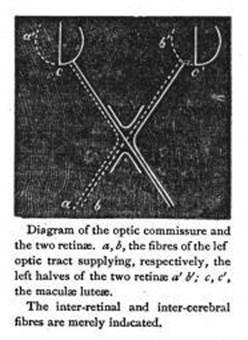
In our own case,
the point to which we desire to call special attention is the rigid optical examination of the region between the
porus options and the macula lutea. The semi-decussation of the nerves at the chiasm being admitted, it
would naturally be supposed that the fibres a from the left tract would supply the left retina from the porus
opticus toward a' ; but our examination shows that it supplies less than this, viz.: only that part of the retina
from c to a' ; while in the right eye the fibres b, instead of supplying the retina from the porus
opticus to b',
supply more than this, viz.: that part of the retina from c' to b'. In other words the fibres a
and b of the left
optic tract supply, mathematically, the left halves of the two retinae from c to a' and c' to
b', and the right
tract the right halves. As to the cerebral seat of the sense of vision, the amount and depth of the injury to
the brain are too uncertain, perhaps, to warrant us in venturing on any speculations us to its locality."