Journal : The Philadelphia Photographer ; vol. xxiii, no. 275.
New York : Edward L. Wilson, June 5, 1886.
Description : 340-341 p. ; ill.: 2 phots., in-text engrs. ; 24 cm.
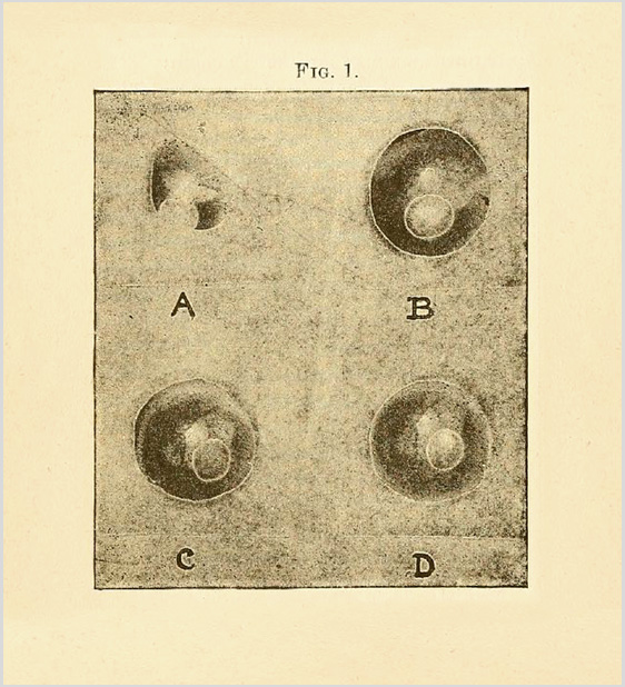
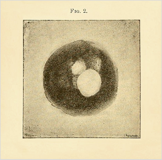
Photographs : two in-text phototypes (Meisenbach process) of the fundus.
Photographers : authors, Jackman & Webster.
Subject : Retina — Fundus photography.
Notes :
After considerable practice, and numerous experiments, we have at last succeeded in obtaining a very fair photograph of the blind spot and a few of the larger bloodvessels of the living human retina. — Jackman & Webster, p. 340. »»:
Jackman and Webster were first to publish photographs of the living human retina, a significant achievement in the history of medical photography. Although some historians try to credit this technical breakthrough to Lucien Howe, Professor of Ophthalmology at the University of Buffalo, and his technical assistant Elmer Starr, their work was published one year later and was illustrated only by drawings. Earlier attempts at photographing the fundus—by adapting a version of the Helmholtz ophthalmoscopic mirror to a camera—were made by a number of scientists, notably, Richard Liebreich (1858), Henry Noyes (1862), A. M. Rosebrugh (1865), Henri Dor (1884), Guiseppe Albertotti (1884); and less notably by B. J. Jeffries (1869), O. F. Wadsworth (1880), Gustav Panel (1887), Charles A. Oliver & Wharton Sinkler (unpublished), and others. These pioneers were constrained by many technical problems, especially by the long exposure time of the emulsions that were available to them. Photographing within the red spectrum of retinal tissue was another intractable problem, as well as distortions and glare from the corneal reflex, involuntary eye movement, and fear of damaging the retina by a blistering focal light source. Jackman and Webster found success with a Westminster ophthalmoscope manufactured by Picard and Curry, and an Argand gas-burner used to illuminate the interior of the eye. Extra sensitive emulsion plates were supplied by Samuel Fry & Co. and reduced exposure time to 2-1/2 minutes, yet with all these technical advances, their procedure still yielded only dim and unremarkable images. The images were degraded further by the cheap Meisenbach process that was used to reproduce the photographs for the journal. Even so, their paper reinvigorated the search for a way to precisely photograph, in vivo, the diseases of the fundus.