London : J. & J. Churchill, 1874.
Description : [4 l.] p., [1]-108 p., [12 l.] pl., [1]-108 p. ; ill.: 8 phot., 2 lith., 2 chrom., in-text engrs. ; 22.3 cm.
Photographs : collotypes on printed leaves.
Photographers : varia.
Subject : Bone — Osteitis, periostitis ; syphilis.
Notes :
Where the ulcer has been deeper round the circumference, the destruction is in a circular or ringlike form, and hence the peculiar appearance of some of the skulls in our museums. There was a very good and characteristic specimen in Parkstreet Museum, now belonging to the Belfast College; and through the kindness of Dr. Cuming, who got photographs taken for me, I am enabled to exhibit them.—Page 67.

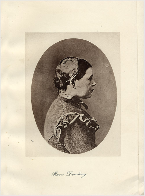
The monograph is divided into seven lectures including one titled, The yellow tubercle, Hamilton's muddling term for the gumma of tertiary stage syphilis. Except for one clinical portrait, the photographic plates represent specimens of diseased bone.
