Leipzig : Veit & Comp., 1878.
Journal : Archiv für Anatomie und Entwickelungsgeschichte ; supplemental volume.
Description : 53–82 p., [3 l.] pl. ; illus: 11 phot. figs. ; 23.5 cm.
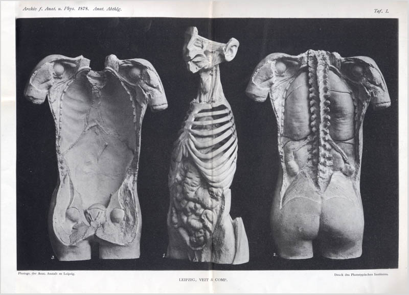
Photographs : composite collotypes on printed leaves, each with views of plaster models.
Subject : Viscera — Morphology ; anatomical models.
Notes :
Because Wilhelm His was first and foremost a morphologist, a preeminent scholar on the anatomical form, he used every technical means possible to transpose the object of his research into a clear graphic on the page, a table of precise measurements, or an accurate three-dimensional model. What was unavailable, he improvised or invented. He was a master draftsman and a superb photographer. The camera was indispensable to his work, but so too were the magic lantern, the stains and solutions that he perfected to color and fix tissue, and the wax-plate method of building a model (his invention). By means of the instrument most closely associated with his name, the His microtome, he was able to observe and capture the cascade of tissue bodies morphing within the developing embryo, and thereby refute Haeckel's recapitulation theory. The "embryograph" was his improvement over the camera lucida, taking a Hartnack microscope and retrofitting it with a drawing prism and an adjustable photographic objective. The most important of his observations were published in the revelatory "Anatomie der menschlichen Embryonen" (1880-1885), regarded as one of the foundational works in the history of embryology.
For this paper on the morphology of the major organs, Wilhelm His introduced the first of a series of superior anatomical models that soon found homes in the anatomical museums and medical schools around the world. Partnering with the sculptor Franz Josef Steger (1845-1938), Dr. His began by injecting a cadaver with five to ten litres of a chromic acid solution under pressure. After the viscera had sufficiently hardened, the limbs were reduced to stumps and the body was propped up and completely encased in plaster. This method allowed the deconstruction of the torso by layers, sawing from different angles and making casts as the work progressed. Strings attached to the skin, with their ends emerging from the plaster shell, provided a guide for the saw and kept the skin from detaching from the forms during sectioning. Eight bodies were used in this report, four male and four female.