Journal : Annals of gynaecology and pediatry ; vol. i.
Boston : Rockwell and Churchill, 1888.
Description : [4 l.] pl., 537-539 p. ; ill.: 8 phot. figs. ; 23 cm.
Photograph : 8 photoengravings on 4 printed leafs.
Subject : Uterus — Prolapse of.
Notes :
Photo captions:
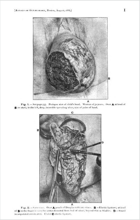
Fig. 1. — See page 537. Prolapse size of child's head. Woman of 70 years. Over A at level of B os uteri;
to the left, deep, incuable spreading ulcer, size of palm of hand.
Fig. 2. — Same case. Over A pouch of Douglas with inte4stines. B — Elastic ligature; at level of
B on the finger is seen the ureter dissected from bed of ulcer; beyond this is bladder. D — Sound in
amputated cervix uteri. Under C elastic ligature.
Fig. 3. — Same case. Douglas' pouch and bladder pushed up, and wound united with silk. Whole mass was then returned
into pelvis; perfect recovery.
Fig. 4. — A — Prolapse of hypertrophied cervix. B — Hypertrophic elongation of post. lip.
Page 538.
Fig. 5. — Complete prolapse. A — In centre of field ulcer of cervix and adjacent parts. Page 538.
Fig. 6. — Same case. Shows ulcer at level of A. At level of B ruptured perinaeum. Amputation of cervix;
ant. colporrhaphy; pernaeorrhaphy. Complete recovery.
Fig. 7. — Vesico-cervical fistula (page 539) in middle line. At level of A fistula. At level of B sound
in os uteri. Cervix torn and ant. lip rolled up.
Fig 8. — Same case. In middle line at level of A repair of fistula. At level of B end of black rubber
plug in uterine canal. C — Speculum of lead pipe. Fifth operation; recovery.

